This is a detailed guide: how to reach a diagnosis in a case of sinus or a case of fistula.
Medical History:
- Duration and mode of onset: are present since birth, (a) Conyenitai sinuses and preauricular sinus and bronchial fistula. (b) Acquired eases usually follow an infective, ulcerative, obstructive or malignant lesion, e.g. chronic osteo-myelitis, tuberculous sinuses, faecal fistula. and fistula-in-ano.
- Discharge: Whether clear mucus (e.g. branchial and salivary fistula) , pus (e.g. chronic osteomyelitis, tuberculosis and stitch sinuses), urine (renal, ureteric, vesical and urethral fistula) , bilious fluid (biliary and duodenal fistula!). flatus and faecal material (faecal fistula) or bone chips (osteomyelitis). In-quire also about the amount of the discharge ; whether scanty or profuse. continuous or intermittent.
LOCAL EXAMINATION
- Number: One opening is usually present but multiple openings commonly occur in osteomyelitis, tuberculosis. actino-mycosis. fistula-in-ano and urethral fistula (watering-can perineum).
- Site: Congenital sinuses and fistulae have characteristic sites, e.g. preauricular sinus (at the root of the helix or on the tragus), branchial fistula (at the lower third of the anterior border of the sternomastoid) and pilonidal sinus (in the midline over the coccyx). In acquired cases, the site is helpful in the diagnosis as in salivary fistulae (face), osteomyelitis (over the ends of long bones), tuberculous sinuses (neck), urinary fistulae (loin, perineum), etc.
- Opening: Exuberant granulation tissue at the orifice is suspicious of a foreign body in the depth, e.g. ligature material, sequestrum, drainage tube or bullet. In tuberculous sinuses the opening resembles a tuberculous ulcer, having a thin blue under-mined edge.
- Discharge: Note its amount, character and smell. The discharge from urinary, biliary and faecal fistulae is usually copious and easily identified. Foul faecal-smelling pus discharg-ed from an abdominal wound should be differentiated from faecal fistula by the absence of gas bubbles or flatus in the dis-charge. The causative organisms of a purulent discharge may be suspected from the physical characteristics of the discharge, e.g the thin yellow discharge, sometimes blood-stained, of streptococcal infection ; the thick whitish creamy pus of staphy-lococcal infection ; the bluish-green pus of infection by Ps. pyo-(-yam% ; and the dark foul-smelling pus of B. soli and B. proteus infection. Actinoinveotie pus is watery and contains sulphur gtamiles which settle to the bottom when the pus is shaken with water in a test-tube.
- Surrounding skin: Note the presence of scarring, puck-ering, dermatitis, excoriation, pigmentation, etc.
- Deeper tissues a) Palpate the surrounding tissues for warmth, tenderness, induration, palpable track or lump. b) Try to establish the origin of the sinus or fistula by examining all important structures in the affected area, e.g. bones, joints, lymph nodes, body cavities or hollow viscera.
- Track: a) Test the mobility of the sinus or track on the under-lying structures and try to determine to which structure it is fixed, e.g. thickened bone, tuberculous glands, urethra, testis, 'etc. b) Insert a blunt probe gently into the sinus and note the length and direction of the track ; the presence of a sequestrum or foreign body ; whether the end of the probe enters a bony cavity or a hollow viscus ; and whether fresh discharge comes out on withdrawal of the probe.
- Lymph nodes: Examine the lymph nodes draining the area for enlargement, consistency, mobility and tenderness.
SPECIAL INVESTIGATIONS
1. Examination of the discharge chemically, microscopically and bacteriologically.
2. Radiography :
- a) Plain films reveal any bony lesions and may show a sequestrum or radio-opaque foreign body.
- b) Fistulography by the injection of lipiodol into the track demonstrates the course and relations of the track and may 'determine its cause and origin.
- c) Contrast rudiography is useful in fistule arising in hollow viscera (e.g. bowel, urinary tract strate the affected viscus, the site of the internal opening and the presence of any distal obstruction.
3. Biopsy is helpful in obscure cases,particularly when specific infection or malignancy is suspected.
Differential diagnosis of Sinuses:
- Congenital sinuses occur at characteristic sites, e.g. pre-auricular sinus in the face and pilonidal sinus over the sacrococcygeal joint. They often remain dry and symptomless until suppuration occurs.
- Stitch sinus is a common complication of sutured wounds. The sinus lies in the line of the incision and follows rupture of a stitch abscess. The track extends down to the offending knot and the discharge continues until the knot is extruded spontaneously or removed by means of a curet-tage spoon.
- Osteomyelitis sinuses are often multiple and adher-ent to the underlying bone which is thickened and tender. Pouting granulation tissue at the mouth of the sinus suggests the presence of a sequestrurn Which imparts a grating sensation on Probing the track. X-ray examination shows the sequestrum lying in a cavity surrounded by an involucrum.
- Empyema sinus: cormmunicates with a chronic empyema cavity through a track in the chest wall. It is associathd with toxamia, amemia and clubbing of the fingers, and acute exacerbations are common with fever, dyspnea, cough and chest pain. Exploration with a probe will show whether the sinus leads to a rib, pleural recess or lung. X-ray examination shows the opacity of the empyerna and may reveal foreign bodies or underlying lung disease. A Iipiodal pleuro-gram demonstrates the size of the empyema cavity and the site of any bronchial communi-cation. A few crystals of Sud-an III added to the lipiodol will appear in the sputum if a bronchopleural fistula is pre-sent.
- Tuberculous sinuses follow bursting or incision of tuberculous cold abscesses. The sinuses are of-ten multiple and surrounded by tuberculous ulceration of the skin and multiple unhealthy scars. The opening has an irre-gular outline with undermined edges and pale anaemic granu-lations. The discharge is thin and watery with flakes of fibrin arid necrotic material. Examination reveals the causative lesion which may underlie the sinus (e.g. tuberculous lvmphadenitis, osteitis and epididymitis) or may be situated far away (e.g. Pott's disease).
- Actinomycotic sinuses are surrounded by a hard indurated mass with rather well-defined edges. The discharge is watery and contains sulphur granules which consist of colonies of the fungus. They are best demonstrated by expressing a little of tho discharge into a test tube half-full of water and shaking the corked tube vigorously the sulphur granules will sink to the bottom of the test-tube.
- Madura foot is a fungus disease allied to actinomycosis. It is endemic in India and several other tropical countries but is rarely seen in Egypt. The disease starts in the subcutaneous tissues and soon involves the deeper structures including the bones and joints. It produces multiple nodules which break down to form sinuses discharging an oily or viscid pus containing granules of the fungus.
Differential diagnosis of Fistulae
- Congenital fistulae occur ar characteristic sites e.g thyroglossal fistula in the midline of the neck , branchial fistula at the anterior border of the sternomastoid and urachal fistula at the umbilicus.
- Faecal fistula presents as a discharging sinus on the abdominal wall, often associated with excoriation of the skin and, sometimes, with dehydration and loss of weight. The discharge from a duodenal or Jejunaal fistula is bile-stained and contains undigested food. When the ileum or cecum is involved, the discharge is fluid faecal matter and when the fistula is connected with the colon, it consists of solid or semi-solid feces. When the leak is small, it may be difficult to distinguish a faecal discharge from fculent pus. The passage of large bubbles of flatus indicates that there is a communication with the bowel and this is confirmed by the appearance of black granules in the discharge after the administration of charcoal by mouth. A barium meal or enema reveals the site of the leak, the length of the track and the presence of any obstruction. If the fistula cannot be demonstrated, the track may be injected with lipiodol to outline its length and direction.
- Umbilical fistulae: The umbilicus, being a central abdominal scar, forms a weak spot in the abdominal wall towards which any fistulous leak is liable to track to open at the surface. Three types of fistulae are encountered, a) Fcecal fistula due to patent vitello-intestinal duct, tuberculous peritonitis or malignant ulceration of the transverse colon. b) Urinary fistula due to patent urachus or infected urachal diverticulum. c) Biliary fistula due to perforation of an inflamed gall-bladder.
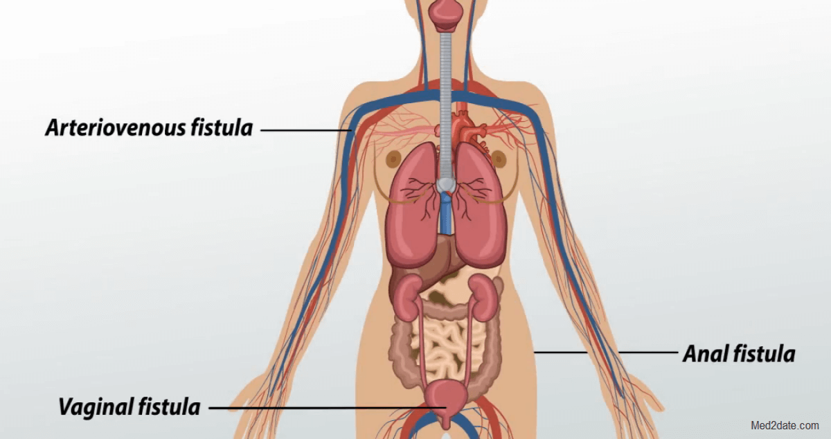
- Anal fistula is intimately related to the anal canal and anus and usually follows bursting or incision of anorectal abscess. It should be differentiated from other sinuses in theperineum, e.g. urethral fistula and pilonidal sinus, by thorough examination, probing and, if necessary, by fistulography, The track may lie entirely under the skin or mucous mem-brane(subcutaneous and submucous fistuice), may course between the sphineteric muscles (intermuscular fistula) or may extend above the anorectal ring (supramuscular fistula). The track may have one external opening (simple fistula) or may have side tracks and multiple external openings (branching or compound fistula). Occasionally, the fistula is merely a sinus track with one opening only (blind fistula).
- Urinary fistula may occur in relation to the kid-ney, ureter, bladder or ure-thra. In the perineum, urin-ary fistulae commonly occur as a complication of gonor-rha)a, bilharziasis , rupture or stricture of the urethra. In bilharzial cases, which are very common, the track usually arises in the bulbous uretiaa, and discharges externally through multiple openings (waiering-can perineum) . In the abdomen, most cases follow operations on the kidneys, ureters or bladder.