📋 Key Information Summary
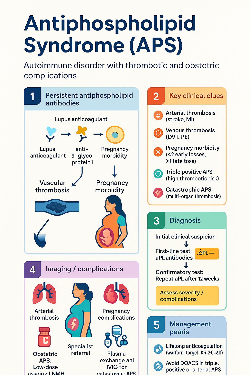
- Antiphospholipid syndrome (APS) is an autoimmune thromboinflammatory disorder defined by persistent antiphospholipid antibodies (aPL) and clinical criteria of vascular thrombosis or pregnancy morbidity.
- Diagnosis requires meeting the updated 2023 ACR/EULAR criteria, which supersede the 2006 Sydney criteria. Classification mandates at least one clinical and one laboratory criterion.
- Laboratory criteria require positive aPL (lupus anticoagulant, anticardiolipin, or anti-β2-glycoprotein I antibodies) on two or more occasions at least 12 weeks apart.
- Clinical manifestations include arterial (stroke, MI) or venous thrombosis (DVT, PE) and specific pregnancy morbidity (e.g., ≥3 early losses, ≥1 late loss, eclampsia).
- Triple positive APS (positive for all three aPL) carries the highest thrombotic risk and requires more aggressive anticoagulation.
- Catastrophic APS (CAPS) is a rare, life-threatening form with rapid-onset multi-organ thrombosis. Treatment involves anticoagulation, corticosteroids, plasma exchange, and/or IVIG.
- Lifelong anticoagulation with warfarin (target INR 2.0-3.0) is standard for thrombotic APS. Direct oral anticoagulants (DOACs) are generally not recommended, especially in arterial or triple positive APS.
- For obstetric APS, management involves low-dose aspirin plus prophylactic low-molecular-weight heparin (LMWH) throughout pregnancy and the postpartum period.
- Investigations include solid-phase assays for aPL and a functional lupus anticoagulant test. Thrombophilia screens should be interpreted in context.
- Aboriginal and Torres Strait Islander peoples experience higher rates of autoimmune disease and may face barriers to specialist care, impacting diagnosis and management.
Introduction & Australian Epidemiology
Antiphospholipid syndrome (APS) is a systemic autoimmune disorder characterised by the presence of circulating antiphospholipid antibodies (aPL) associated with a high risk of vascular thrombosis and pregnancy complications. It may occur in isolation (primary APS) or in association with other autoimmune diseases, most commonly systemic lupus erythematosus (SLE).
In Australia, the epidemiology mirrors that of other developed nations. APS is a significant cause of acquired thrombophilia and recurrent pregnancy loss. The incidence is estimated at 5-10 new cases per 100,000 population per year, with a prevalence of 40-50 per 100,000. It predominantly affects women, with a female-to-male ratio of approximately 4:1. The disorder has considerable impact on healthcare utilisation due to the need for long-term anticoagulation and monitoring.
Diagnostic Criteria: Sydney 2006 & ACR/EULAR 2023
The diagnosis of APS has evolved. The revised Sydney criteria (2006) formed the standard for many years but have now been replaced by the 2023 ACR/EULAR classification criteria, which offer improved specificity and incorporate newer clinical and laboratory features.
ACR/EULAR 2023 Classification Criteria
An individual is classified as having APS if they meet at least one clinical criterion and at least one laboratory criterion, with the laboratory test being positive on two or more occasions at least 12 weeks apart. Entry criterion: at least one positive aPL test within 3 years of a clinical criterion.
| Domain | Criterion | Points |
|---|---|---|
| Clinical |
Macrovascular venous thrombosis: Image-proven or objectively confirmed superficial or deep venous thrombosis (excluding superficial venous thrombosis in varicose veins). Macrovascular arterial thrombosis: Image-proven or objectively confirmed arterial thrombosis in any tissue/organ (excluding liver, portal, splenic, mesenteric veins). Microvascular: Suspected or proven nephropathy (aPL-associated nephropathy), adrenal haemorrhage, myocardial microthrombosis, or livedo racemosa. Pregnancy morbidity: ≥3 consecutive pre-fetal/early neonatal deaths, ≥1 fetal death after 10 weeks, or ≥1 premature birth before 34 weeks due to eclampsia/preeclampsia/placental insufficiency. |
Up to 5 points per domain (weighted) |
| Laboratory |
Lupus anticoagulant (LAC): Positive according to ISTH guidelines. Anti-cardiolipin (aCL) IgG/M: >40 GPL/MPL or >99th percentile. Anti-β2-glycoprotein I (aβ2GPI) IgG/M: >99th percentile. |
Up to 7 points (weighted, with higher scores for positivity in multiple tests/medium-high titres) |
Arterial & Venous Thrombosis
Thrombosis is the hallmark clinical manifestation of APS. It can occur in any vascular bed and may be the first presentation.
- Most common manifestation.
- Deep vein thrombosis (DVT) of the lower limbs is classic.
- Pulmonary embolism (PE) is a frequent and serious complication.
- Unusual site thrombosis: cerebral venous sinus thrombosis, hepatic/portal veins, retinal veins, adrenal veins.
- Ischaemic stroke is the most common arterial event, often in patients <50 years.
- Myocardial infarction, peripheral arterial occlusion.
- Amaurosis fugax, retinal artery occlusion.
- Associated with higher morbidity and mortality than venous events.
Pregnancy Morbidity
APS is a significant cause of recurrent pregnancy loss and obstetric complications. The mechanism involves placental thrombosis and inflammation.
Obstetric Criteria (ACR/EULAR 2023)
- ≥3 consecutive spontaneous abortions before week 10 of pregnancy (with maternal anatomical/hormonal and paternal/maternal chromosomal causes excluded).
- ≥1 unexplained death of a morphologically normal fetus at or beyond week 10.
- ≥1 premature birth before week 34 due to eclampsia, severe preeclampsia, or recognised features of placental insufficiency.
Associated Complications
Beyond pregnancy loss, APS increases the risk of preeclampsia, eclampsia, placental abruption, fetal growth restriction, and preterm labour.
Anticoagulation: Warfarin vs. DOACs
Anticoagulation is the cornerstone of APS management for thrombotic events, but the choice of agent is critical.
Warfarin (First-line for thrombotic APS)
Direct Oral Anticoagulants (DOACs) – Role in APS
Consideration of a DOAC (e.g., apixaban) may be restricted to a very limited subset: patients with single positive (low-titre aCL or aβ2GPI only, LAC negative) venous-only APS who have contraindications or extreme difficulty with warfarin management, after discussion with a specialist.
Obstetric Anticoagulation
For pregnancy, warfarin is teratogenic. Management involves:
- Pre-conception: Switch from warfarin to LMWH.
- During pregnancy: Low-dose aspirin (75-100 mg daily) plus prophylactic or intermediate-dose LMWH (e.g., enoxaparin 40 mg SC daily).
- Postpartum: Continue anticoagulation (warfarin or LMWH) for at least 6 weeks, often lifelong if thrombotic history.
Triple Positive APS
Triple positive APS is defined by the persistent presence of all three laboratory markers: lupus anticoagulant (LAC), anticardiolipin antibodies (aCL), and anti-β2-glycoprotein I antibodies (aβ2GPI). This profile represents the highest risk category within the APS spectrum.
- Annual thrombosis risk estimated at 5-10%, even on anticoagulation.
- Highest risk of recurrent thrombosis, especially arterial.
- DOACs are contraindicated due to high failure rates.
- Requires lifelong warfarin with strict INR control (target 2.0-3.0).
Management is inherently specialist-driven. Treatment beyond standard anticoagulation (e.g., addition of hydroxychloroquine, statins) is an area of active research but not yet standard of care.
Catastrophic APS (CAPS)
Diagnostic Criteria (Preliminary Criteria for CAPS)
- Evidence of involvement of ≥3 organs, systems, and/or tissues.
- Development of manifestations simultaneously or in less than a week.
- Confirmation by histopathology of small vessel occlusion in at least one organ/tissue.
- Laboratory confirmation of the presence of aPL (LAC and/or aCL/aβ2GPI).
Definite CAPS: All four criteria. Probable CAPS: All four criteria but only 2 organs; or criteria 1, 2, and 4; or criteria 1, 3, 4 and a 1-week delay in a third organ.
Management (Requires ICU admission)
Treatment is multimodal and must be initiated immediately:
- Anticoagulation: Immediate intravenous heparin, bridging to long-term warfarin.
- Corticosteroids: High-dose methylprednisolone pulse (1g IV daily for 3 days) followed by maintenance.
- Plasma Exchange (PLEX): Often first-line adjunct to remove pathogenic antibodies and cytokines.
- Intravenous Immunoglobulin (IVIG): 2g/kg divided over 2-5 days, often used in combination or if PLEX unavailable.
- Rituximab/Complement Inhibitors: Considered in refractory cases.
Investigations
Diagnosis relies on specific laboratory tests for aPL. Testing should be performed in the context of a qualifying clinical event and repeated after 12 weeks for confirmation.
Special Populations
ATSI Health Considerations
While specific prevalence data for APS in Aboriginal and Torres Strait Islander populations is limited, systemic autoimmune diseases like SLE are more common and often more severe. Key considerations for equitable care include:
📚 References
- 1. Barbhaiya M, Zuily S, Naden R, et al. The 2023 ACR/EULAR Antiphospholipid Syndrome Classification Criteria. Arthritis Rheumatol. 2023;75(10):1687-1702.
- 2. Miyakis S, Lockshin MD, Atsumi T, et al. International consensus statement on an update of the classification criteria for definite antiphospholipid syndrome (APS). J Thromb Haemost. 2006;4(2):295-306.
- 3. Cohen H, Efthymiou M, Isenberg DA. Antiphospholipid antibodies and the antiphospholipid syndrome. In: Textbook of Autoimmune Rheumatic Diseases. 2nd ed. Oxford University Press; 2022.
- 4. Ortel TL, Neumann I, Ageno W, et al. American Society of Hematology 2020 guidelines for management of venous thromboembolism: treatment of deep vein thrombosis and pulmonary embolism. Blood Adv. 2020;4(19):4693-4738.
- 5. Tektonidou MG, Andreoli L, Limper M, et al. EULAR recommendations for the management of antiphospholipid syndrome in adults. Ann Rheum Dis. 2019;78(10):1296-1304.
- 6. Cervera R, Rodríguez-Pintó I, Espinosa G. The diagnosis and clinical management of the catastrophic antiphospholipid syndrome: A comprehensive review. J Autoimmun. 2018;92:1-11.
- 7. Woller SC, Stevens SM, Kaplan D, et al. Apixaban compared with warfarin to prevent thrombosis in thrombotic antiphospholipid syndrome: a randomized trial. Blood Adv. 2022;6(6):1661-1670.
- 8. Pengo V, Denas G, Zoppellaro G, et al. Rivaroxaban vs warfarin in high-risk patients with antiphospholipid syndrome (TRAPS): a randomized trial. Blood. 2018;132(13):1365-1371.
- 9. Australian Institute of Health and Welfare (AIHW). Aboriginal and Torres Strait Islander health performance framework. 2023.
- 10. Royal Australian College of General Practitioners (RACGP). Management of venous thromboembolism in primary care. 2021.
- 11. Australasian Society of Clinical Immunology and Allergy (ASCIA). Antiphospholipid syndrome - clinical overview. 2023.