Bacterial Morphology
Bacterial Size
Bacterial Shape and Arrangement
- Diplococci: pairs of cells, e.g. Neisseria.
- Irregular grape-like clusters, e.g. staphylococci.
- Chains of four or more, e.g. streptococci.
Staining Characteristics
Bacterial Ultra-Structures and their Functions
 |
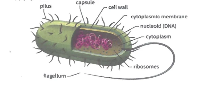
| Fig. 1: schematic presentation of bacterial cell |
Cytoplasm
A few morphologically distinct components can be found within the cytoplasm:
- Nucleoid: Genetic information of a bacterial cell is contained in a single circular molecule of double-stranded DNA, which constitutes the bacterial chromosome. It is 1 mm long and is packed into a supercoiled state inside the cell.
- Plasmids: In many bacteria, additional genetic information is contained on plasmids which are small circular extrachromosomal DNA molecules that can replicate independently of the chromosome.
- Ribosomes: They are the site of protein synthesis in the cell. Ribosomes consist of protein and RNA. Prokaryotic ribosomes have a sedimentation constant of 70S, smaller than the 80S ribosomes of eukaryotes. This difference makes bacterial ribosomes a selective target for antibiotic action.
- Inclusion granules: These are granules of nutrient materials, usually phosphates, sulphur, carbohydrates and lipids. Energy reserves are usually stored as glycogen, starch or poly-B-hydroxybutyrate. Phosphate is stored in metachromatic or volutin granules, which are used for synthesis of ATP.
- Mesosomes: These are complex invagination of the cytoplasmic membrane. They are involved in cell division and sporulation. They also have a function analogous to the mitochondria in eukaryotes providing a membranous support for respiratory enzymes.
Cytoplasmic membrane
In bacteria, as in other cells, the protoplast is limited externally by a thin elastic cytoplasmic membrane. It is a phospholipid protein bilayer similar to that of eukaryotic cells except that, in bacteria, it lacks sterols.
It has the following functions:
- Selective transport: In bacteria, molecules move across the cytoplasmic membrane by simple diffusion, facilitated diffusion and active transport.
- Excretion of extracellular enzymes:
- Hydrolytic enzymes: which digest large food molecules into subunits small enough to penetrate the cytoplasmic membrane.
- Enzymes used to destroy harmful chemicals, such as antibiotics, e.g. penicillin-degrading enzymes.
- Respiration: The respiratory enzymes are located in the cytoplasmic membrane, which is thus a functional analogue of the mitochondria in eukaryotes.
- Cell wall biosynthesis: The cytoplasmic membrane is the site of:
- The enzymes of cell wall biosynthesis.
- The carrier lipids on which the subunits of the cell wall are assembled.
- Reproduction: A specific protein in the membrane attaches to the DNA and separates the duplicated chromosomes from each other. In addition, a septum is formed by the cytoplasmic membrane to separate the cytoplasm of the two daughter cells.
- Chemotactic system: Attractants and repellants bind to specific receptors in the cytoplasmic membrane and send signals to the cell's interior. The cell then responds to the surface message.
Cell Wall
The bacterial cell wall is the structure that immediately surrounds the cytoplasmic membrane. It is 10-25 nm thick, strong and relatively rigid, thogh having some elasticity.
Structure of the cell wall:
The cell wall of bacteria is a complex structure. Its impressive strength is primarily due to peptidoglycan (synonym: murein or mucopeptide). Peptidoglycan is a complex polymer consisting of N-acetylglucosamine (NAG) and N-acetylmuramic acid (NAM) that is unique to bacteria. A set of identical tetrapeptide side chains are attached to NAM.
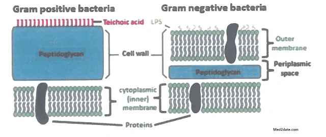
Besides peptidoglycan, additional components in the cell wall divide bacteria into Gram-positive and Gram-negative (Fig. 2).
Gram-positive cell wall is composed of:
- Peptidoglycan layer: Thick, composed of up to 40 sheets, comprising up to 50% of the cell wall material. Despite the thickness of peptidoglycan, chemicals can readily pass through.
- Teichoic acids: Polymers of ribitol or glycerol phosphate. They are found in the cell wall of most Gram-positive bacteria. Teichoic acids and cell wall associated proteins are the major surface antigens of the Gram-positive bacteria.
Gram-negative cell wall is composed of:
- Peptidoglycan layer: Much thinner, composed of only one or two sheets comprising 5-10% of the cell wall material.
- Outer membrane: Phospholipid protein bilayer present external to the peptidoglycan layer. The outer surface of the lipid bilayer is composed of molecules of lipopolysaccharide (LPS) which consists of a complex lipid called lipid A chemically linked to polysaccharides. Lipid A of the LPS forms the endotoxin of the Gram-negative bacteria, while polysaccharides are the outermost molecules of the cell wall and are major surface antigens of the Gram-negative bacterial cell (somatic or O antigen).
Functions of the cell wall
- It maintains the characteristic shape of the bacterium.
- It supports the weak cytoplasmic membrane against the high internal osmotic pressure of the protoplasm (5-25 atm.).
- It plays an important role in cell division.
- It is responsible for the staining affinity of the organism.
Wall deficient variants
- "L" stands for Lister Institute in London, where they were first discovered.
- "L" forms may develop from cells that normally possess cell wall, when they are exposed to hydrolysis by lysozyme or by blocking peptidoglycan biosynthesis with antibiotics, such as penicillin, provided that they are present in an isotonic medium.
- Some L. forms resynthesize their walls once the inducing stimulus is removed. Others, however, permanently lose the capacity to produce a cell wall.
- L. forms may survive antibiotic therapy. Their reversion to the walled state can produce relapses of the overt infection.
Capsule and Related Structures
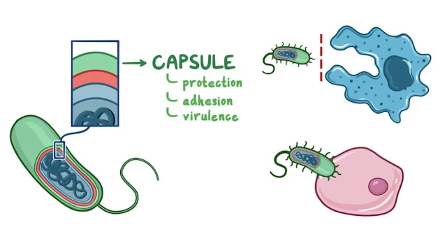
Many bacteria synthesize large amount of extracellular polymer that collects outside the cell wall to form an additional surface layer. This layer is formed only inside the host (in-vivo).- Capsule: It is such a layer that adheres to the surface of the cell and forms a well-defined halo when differentially stained, to be resolved with the light microscope.
- Slime layer: It is a surface layer that is loosely distributed around the cell.
- Glycocalyx: It is a loose meshwork of polysaccharide fibrils extending outwards from the cell.
Functions:
- It protects the cell wall against various kinds of antibacterial agents, e.g. bacteriophages, colicins, complement and lysozymes.
- It protects the bacterial cell from phagocytosis. Hence, the capsule is considered an important virulence factor.
- Some bacteria attach to the target surface by using their capsules or glycocalyx in order to establish infection. For instance, Streptococcus mutans form glycocalyx, with which the bacteria stick to the tooth enamel.
Appendages
Several structures project through the cell wall of bacteria to form surface appendages. The most commonly observed are flagella and pili.
A- Flagella
Many genera of bacteria move by means of flagella.
- Flagella are only 20 nm in diameter, too small to be seen with a light microscope. They can be clearly demonstrated with an electron microscope.
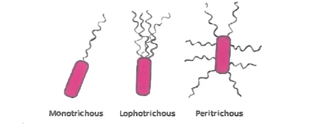
- The location and number of flagella on a cell vary depending on the bacterial species. Organisms may be monotrichous (a single flagellum at one end of the cell), lophotrichous (multiple flagella at one end of the cell), or peritrichous (flagella distributed over the entire cell surface) (Fig. 3).
- Flagella consist of a single type of protein called flagellin, which differs in different bacterial species. Flagellins are highly antigenic (H antigen).
Axial filaments
These structures are composed of two groups of fibers that originate within the opposite ends of the cell and overlap in the middle. Structurally and chemically, the fibers of the axial filaments are similar to flagella and they are sometimes called "endoflagella".
Spirochaetes move by means of these axial filaments. When the cell moves, it rotates around its longitudinal axis and flexes and bends along its length.
B- Pili or fimbriae
Pili (singular: pilus) are protein tubes that extend from the cells. They are shorter and thinner than flagella and can be observed only by the electron microscope. They are composed of structural protein subunits termed pilins.
Functions:
- Adherence: It is the function of the short pili (fimbriae) that occur in great numbers around the cell. They enable bacteria to attach to the surfaces, thus contributing to the establishment of infection i.e. virulence factor. For instance, N. gonorrhoeae withstands the flushing action of urine by adhering to the urethral mucosa.
- Conjugation: A special long pilus called the sex pilus (F pilus) is involved in the transfer of DNA between bacteria, a process known as conjugation.


