📋 Key Information Summary
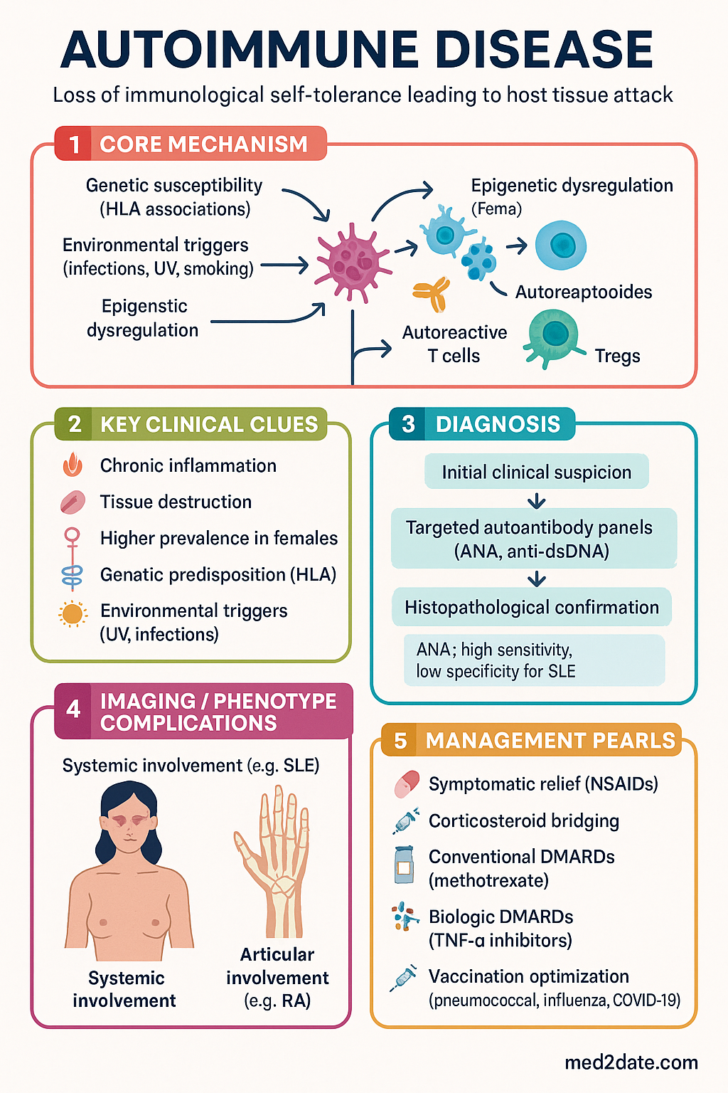
- Autoimmune diseases result from loss of immunological self-tolerance, with the immune system attacking host tissues via autoantibodies, immune complexes, or autoreactive T cells.
- Australia has among the highest autoimmune disease prevalence globally, affecting approximately 5–8% of the population, with higher rates in Aboriginal and Torres Strait Islander communities.
- Classification is divided into organ-specific (e.g., type 1 diabetes, Hashimoto thyroiditis, coeliac disease) and systemic (e.g., systemic lupus erythematosus, rheumatoid arthritis, systemic sclerosis) categories.
- Pathogenesis involves a combination of genetic susceptibility (HLA associations), environmental triggers (infections, UV, smoking), epigenetic dysregulation, and failure of immune checkpoint mechanisms.
- Diagnostic workup centres on clinical assessment, targeted autoantibody panels (ANA, anti-dsDNA, anti-CCP, anti-TPO), inflammatory markers (ESR, CRP), and histopathological confirmation where indicated.
- Anti-nuclear antibody (ANA) testing has high sensitivity but low specificity for SLE; a positive ANA alone is insufficient for diagnosis — correlate with clinical features and specific extractable nuclear antigen (ENA) profiles.
- Management follows a stepwise approach: symptomatic relief (NSAIDs, analgesics) → corticosteroid bridging → conventional DMARDs (methotrexate, hydroxychloroquine, azathioprine) → biologic DMARDs (TNF-α inhibitors, rituximab) → targeted synthetic DMARDs (JAK inhibitors).
- Hydroxychloroquine (Plaquenil®) is PBS-listed for SLE and rheumatoid arthritis; requires baseline and annual ophthalmological screening for retinal toxicity after cumulative doses exceeding 1000 g.
- Methotrexate (Methoblastin®) remains the anchor DMARD for RA; mandatory monitoring includes FBC, LFTs, and renal function every 2–4 weeks initially, then every 2–3 months at stable dose.
- Biologic DMARDs require screening for latent tuberculosis (IGRA or tuberculin skin test), hepatitis B/C serology, and varicella status before initiation.
- Immunosuppressive therapy significantly increases susceptibility to infections — pneumococcal, influenza, and COVID-19 vaccination should be optimised prior to commencing biologics.
- Aboriginal and Torres Strait Islander peoples experience higher disease burden, delayed diagnosis, and poorer outcomes — culturally safe care, community-based follow-up, and PBS co-payment concessions are essential.
- Pre-conception counselling is critical for women of childbearing age, as methotrexate, mycophenolate, and cyclophosphamide are teratogenic; hydroxychloroquine and azathioprine are considered safe in pregnancy.
Introduction & Australian Epidemiology
Autoimmune disease encompasses a broad spectrum of disorders in which the adaptive or innate immune system mounts an inappropriate response against self-antigens, resulting in chronic inflammation and tissue destruction. This failure of immunological self-tolerance may manifest as organ-specific disease — where a single organ is targeted — or as systemic disease with multi-organ involvement.
Australia has one of the highest prevalences of autoimmune disease in the world. An estimated 1.2 million Australians (approximately 5–8% of the population) live with one or more autoimmune conditions. Rheumatoid arthritis alone affects approximately 456,000 Australians, while systemic lupus erythematosus (SLE) disproportionately affects women of childbearing age at a ratio of approximately 9:1 (female to male). Type 1 diabetes, coeliac disease, autoimmune thyroiditis, and inflammatory bowel disease collectively represent the most common organ-specific autoimmune conditions encountered in Australian primary care.
Key epidemiological observations in Australia include:
- Rheumatoid arthritis prevalence is 1.9% in women and 0.8% in men, with higher rates in Aboriginal and Torres Strait Islander populations.
- SLE prevalence in Australia is approximately 45 per 100,000, with significantly higher rates in Indigenous Australians, particularly those of mixed ancestry.
- Type 1 diabetes incidence in Australian children (0–14 years) is approximately 23 per 100,000 per year — among the highest rates globally.
- Coeliac disease affects approximately 1 in 70 Australians, though many remain undiagnosed; active case-finding is recommended in at-risk groups.
- Autoimmune thyroiditis (Hashimoto disease) is the most common autoimmune condition in Australia, with a prevalence of 3–5%.
- Geographic and climatic variations influence disease patterns — UV radiation exposure is a recognised trigger for SLE flares in northern Australia, while vitamin D deficiency in southern latitudes may modulate immune function.
Sex and Gender Considerations
Approximately 78% of individuals affected by autoimmune disease are female. Oestrogen promotes B-cell survival and antibody production, while X-chromosome inactivation mosaicism may contribute to greater immune diversity and self-reactivity. These sex-based differences have significant implications for screening, diagnosis, and management in Australian clinical practice.
Classification & Types
Autoimmune diseases are classified based on the target of immune attack and the breadth of organ involvement. This classification guides diagnostic workup, specialist referral pathways, and therapeutic strategies relevant to Australian practice.
Organ-Specific Autoimmune Diseases
In organ-specific autoimmune diseases, the immune response is directed against antigens confined to a single organ or tissue. These conditions are typically managed by organ-specific specialists in the Australian healthcare system.
| Condition | Target Organ | Key Autoantibodies | Typical Specialist | Australian Prevalence |
|---|---|---|---|---|
| Hashimoto thyroiditis | Thyroid | Anti-TPO, anti-thyroglobulin | Endocrinologist / GP | 3–5% |
| Graves disease | Thyroid | TSH receptor antibodies (TRAb) | Endocrinologist | ~0.5% |
| Type 1 diabetes | Pancreatic β cells | Anti-GAD65, anti-IA-2, anti-ZnT8 | Endocrinologist / Paediatrician | ~0.5% |
| Coeliac disease | Small bowel mucosa | Anti-tTG IgA, anti-endomysial | Gastroenterologist / GP | ~1.4% |
| Autoimmune hepatitis | Liver | ANA, anti-SMA, anti-LKM-1 | Gastroenterologist / Hepatologist | ~24 per 100,000 |
| Primary biliary cholangitis | Intrahepatic bile ducts | Anti-mitochondrial (AMA) | Gastroenterologist / Hepatologist | ~35 per 100,000 |
| Multiple sclerosis | CNS myelin | Oligoclonal bands (CSF) | Neurologist | ~100 per 100,000 |
| Myasthenia gravis | Neuromuscular junction | Anti-AChR, anti-MuSK | Neurologist | ~20 per 100,000 |
| Autoimmune haemolytic anaemia | Red blood cells | Direct Coombs test positive | Haematologist | ~1–3 per 100,000/yr |
| Pemphigus vulgaris | Skin / mucosal epithelium | Anti-desmoglein 1 & 3 | Dermatologist | ~1–5 per 100,000 |
Systemic Autoimmune Diseases
Systemic autoimmune diseases involve immune-mediated damage to multiple organs and tissues, often through circulating autoantibodies, immune complex deposition, or widespread T-cell activation. These are predominantly managed by rheumatologists in collaboration with organ-specific specialists.
| Condition | Organs Affected | Key Autoantibodies | Hallmark Feature | F:M Ratio |
|---|---|---|---|---|
| Systemic lupus erythematosus (SLE) | Skin, joints, kidneys, brain, serosal surfaces | ANA, anti-dsDNA, anti-Smith, anti-Ro/La | Multi-system flares, nephritis | 9:1 |
| Rheumatoid arthritis (RA) | Synovial joints (symmetric) | RF, anti-CCP (ACPA) | Symmetric polyarthritis, erosions | 3:1 |
| Systemic sclerosis (scleroderma) | Skin, lungs, GI tract, vasculature | Anti-Scl-70, anti-centromere, anti-RNA pol III | Skin fibrosis, Raynaud, ILD / PAH | 4:1 |
| Sjögren syndrome | Salivary, lacrimal glands, extraglandular | Anti-Ro (SSA), anti-La (SSB), RF | Sicca symptoms, fatigue | 9:1 |
| Systemic vasculitis (ANCA-associated) | Small-to-medium vessels, kidneys, lungs | c-ANCA (PR3), p-ANCA (MPO) | GPA, MPA, EGPA subtypes | 1:1 |
| Dermatomyositis / Polymyositis | Skeletal muscle, skin (dermatomyositis) | Anti-Jo-1, anti-Mi-2, anti-MDA5 | Proximal myopathy, heliotrope rash | 2:1 |
| Antiphospholipid syndrome | Vascular endothelium (thrombotic) | Anti-cardiolipin, anti-β2GP1, lupus anticoagulant | Recurrent thrombosis, pregnancy loss | 5:1 |
Mixed and Overlap Syndromes
A significant proportion of Australian patients present with features spanning multiple autoimmune categories. Overlap syndromes include:
- Mixed connective tissue disease (MCTD): Features of SLE, systemic sclerosis, polymyositis, and RA with high-titre anti-U1 RNP antibodies.
- Anti-synthetase syndrome: Myositis, interstitial lung disease, mechanic's hands, arthritis, Raynaud phenomenon, with anti-aminoacyl-tRNA synthetase antibodies.
- Primary Sjögren syndrome with RA overlap: Common in clinical practice; requires integrated management of sicca symptoms and inflammatory arthritis.
- Scleroderma-myositis overlap: Skin thickening with proximal muscle weakness; requires combined rheumatology and respiratory monitoring for interstitial lung disease.
Pathogenesis
The pathogenesis of autoimmune disease is multifactorial, involving an interplay between genetic predisposition, environmental triggers, immune dysregulation, and stochastic events. Understanding these mechanisms is essential for rational therapeutic targeting and risk stratification in Australian patients.
Failure of Central and Peripheral Tolerance
Immunological self-tolerance operates at two levels:
- Central tolerance: In the thymus (T cells) and bone marrow (B cells), autoreactive lymphocytes are eliminated through negative selection. Defects in the AIRE (autoimmune regulator) gene impair thymic expression of peripheral tissue antigens, allowing autoreactive T cells to escape into the periphery. AIRE mutations cause autoimmune polyendocrinopathy-candidiasis-ectodermal dystrophy (APECED/APS-1).
- Peripheral tolerance: Autoreactive lymphocytes that escape central deletion are controlled by regulatory T cells (Tregs, CD4+CD25+FoxP3+), anergy (functional unresponsiveness), and activation-induced cell death. Deficiency or dysfunction of Tregs is a hallmark of many autoimmune diseases.
Genetic Susceptibility
Genome-wide association studies (GWAS) have identified over 200 susceptibility loci for autoimmune disease. The HLA region on chromosome 6p21 remains the strongest genetic risk factor for most conditions:
| Disease | HLA Association | Odds Ratio | Non-HLA Genes |
|---|---|---|---|
| Rheumatoid arthritis | HLA-DRB1 (shared epitope) | 3–6 | PTPN22, STAT4, CTLA4 |
| SLE | HLA-DR2, HLA-DR3 | 1.5–3 | IRF5, STAT4, TNFAIP3, complement genes (C2, C4) |
| Type 1 diabetes | HLA-DR3, HLA-DR4, HLA-DQ8 | 6–20 | INS, PTPN22, IL2RA, CTLA4 |
| Coeliac disease | HLA-DQ2, HLA-DQ8 | ~100 (necessary but not sufficient) | SH2B3, IL18RAP, TAGAP |
| Ankylosing spondylitis | HLA-B27 | ~100 | ERAP1, IL23R, IL12B |
| Multiple sclerosis | HLA-DRB1*15:01 | 3 | IL7R, IL2RA, CD58 |
Environmental Triggers
Environmental factors are critical in initiating or exacerbating autoimmune disease in genetically susceptible individuals. Triggers with particular relevance in the Australian context include:
- Ultraviolet radiation: Australia's high UV index (particularly northern latitudes) is a well-established trigger for SLE flares, inducing keratinocyte apoptosis and exposing intracellular autoantigens. UVB also modulates dendritic cell function and promotes type I interferon production.
- Infections (molecular mimicry): Streptococcal pharyngitis triggers rheumatic heart disease; EBV is strongly associated with SLE and MS; Campylobacter jejuni triggers Guillain-Barré syndrome; hepatitis C is linked to cryoglobulinaemic vasculitis. Molecular mimicry occurs when microbial peptides share structural homology with self-antigens.
- Smoking: Cigarette smoking is the strongest modifiable environmental risk factor for seropositive RA (anti-CCP positive), increasing risk 2–3-fold, particularly in HLA-DRB1 shared epitope carriers. Smoking also exacerbates SLE and Graves disease.
- Silica and organic solvents: Occupational silica dust exposure (mining, construction) increases RA and SLE risk — relevant to Australia's resource sector. Organic solvents are associated with systemic sclerosis.
- Vitamin D: Vitamin D deficiency, prevalent in southern Australian states during winter, modulates immune function and has been epidemiologically linked to increased MS and type 1 diabetes incidence. Optimal 25-hydroxyvitamin D levels ≥75 nmol/L are recommended.
- Gut microbiome: Dysbiosis (reduced Firmicutes, increased Bacteroidetes) is implicated in RA, SLE, and IBD. Diet, antibiotic exposure, and geographic factors shape the microbiome in Australian populations.
- Hormonal factors: Oestrogen upregulates B-cell activating factor (BAFF) and promotes class-switch recombination. Pregnancy modulates disease activity (RA improves in pregnancy but SLE may flare). Postpartum flares are common across many autoimmune conditions.
Immunopathological Mechanisms
Tissue damage in autoimmune disease is mediated through four classical mechanisms (Gell & Coombs classification):
The Cytokine Network in Autoimmunity
Cytokine dysregulation is central to autoimmune pathogenesis and provides the molecular basis for biologic DMARD therapy. Key cytokine pathways targeted by current and emerging therapies include:
- TNF-α: Pro-inflammatory cytokine central to RA, IBD, psoriasis, and spondyloarthropathy pathogenesis. Targeted by adalimumab (Humira®), etanercept (Enbrel®), infliximab (Remicade®).
- IL-6: Drives acute-phase response, B-cell differentiation, and Th17 differentiation. Targeted by tocilizumab (Actemra®) and sarilumab (Kevzara®).
- IL-17: Produced by Th17 cells; promotes neutrophil recruitment and tissue inflammation in psoriasis, spondyloarthropathy, and RA. Targeted by secukinumab (Cosentyx®) and ixekizumab (Taltz®).
- IL-23: Drives Th17 cell maintenance and expansion. Targeted by guselkumab (Tremfya®) and risankizumab (Skyrizi®).
- Type I interferons (IFN-α/β): Plasmacytoid dendritic cell overproduction of IFN-α is a hallmark of SLE. Targeted by anifrolumab (Saphnelo®), now TGA-approved in Australia for SLE.
- BAFF (B-cell activating factor): Promotes B-cell survival and is elevated in SLE. Targeted by belimumab (Benlysta®).
- JAK-STAT pathway: Intracellular signalling cascade downstream of multiple cytokine receptors. Targeted by tofacitinib (Xeljanz®), baricitinib (Olumiant®), upadacitinib (Rinvoq®).
Diagnostic Approach
The diagnostic approach to autoimmune disease requires integration of clinical assessment, serological testing, imaging, and histopathology. A systematic approach is essential to avoid diagnostic delay, which remains a significant issue in Australia — average time to diagnosis for conditions such as SLE and systemic sclerosis is 2–6 years from symptom onset.
Clinical Assessment
A thorough history and examination should focus on:
- Symptom pattern: Duration, distribution (symmetric vs. asymmetric), chronicity, and systemic features (fatigue, weight loss, fevers, night sweats).
- Organ-specific enquiry: Joint pain/swelling, skin rashes, Raynaud phenomenon, sicca symptoms, oral ulcers, alopecia, dysphagia, chest pain (serositis), visual changes, neurological symptoms.
- Family history: Autoimmune diseases cluster in families; ~30% of patients with one autoimmune condition will develop another (polyautoimmunity).
- Medication history: Drug-induced lupus (hydralazine, procainamide, isoniazid, minocycline) must be excluded.
- Reproductive history: Recurrent miscarriage may indicate antiphospholipid syndrome.
- Occupational and environmental exposures: Smoking, silica dust, solvents, UV exposure.
Laboratory Investigations
Tier 1 — Screening (GP-accessible, MBS-rebatable)
Tier 2 — Specialist-directed
Histopathology
Tissue biopsy remains the gold standard for diagnosing and staging several autoimmune conditions:
- Renal biopsy: ISN/RPS classification of lupus nephritis (Class I–VI) guides treatment intensity. Repeat biopsy warranted for suspected transformation or treatment failure.
- Skin biopsy: Direct immunofluorescence (lupus band test), histopathology for vasculitis, pemphigus/pemphigoid subtyping.
- Small bowel biopsy: Villous atrophy, crypt hyperplasia, intraepithelial lymphocytosis (Marsh classification) in coeliac disease — though diagnosis may be made without biopsy in children with tTG IgA >10× ULN and positive EMA.
- Salivary gland biopsy: Minor salivary gland biopsy (focus score ≥1) supports Sjögren syndrome diagnosis when serology is equivocal.
- Liver biopsy: Interface hepatitis with plasma cell infiltrate in autoimmune hepatitis; staging guides treatment decisions.
Classification Criteria
Validated classification criteria assist in standardising diagnosis and facilitating clinical trial recruitment. Key criteria sets used in Australian practice include:
- SLE: 2019 EULAR/ACR classification criteria (ANA ≥1:80 entry criterion, then additive weighted domains; ≥10 points = classified as SLE).
- RA: 2010 ACR/EULAR classification criteria (joint involvement, serology, acute-phase reactants, symptom duration; ≥6/10 points = classified as RA).
- Systemic sclerosis: 2013 ACR/EULAR criteria (skin thickening of fingers extending proximal to MCPs as sufficient criterion, or ≥9 points from 7 items).
- ANCA-associated vasculitis: 2022 ACR/EULAR classification criteria for GPA, MPA, and EGPA.
- Sjögren syndrome: 2016 ACR/EULAR criteria (anti-Ro positivity, focal lymphocytic sialadenitis, ocular staining score, Schirmer test, salivary flow; ≥4 points).
Referral Pathways in Australia
| Clinical Scenario | Referral | Urgency |
|---|---|---|
| Inflammatory polyarthritis (suspected RA) | Rheumatologist | Within 6 weeks of symptom onset (treat-to-target) |
| Suspected SLE with renal involvement | Rheumatologist + Nephrologist | Urgent (days) |
| Suspected ANCA vasculitis | Rheumatologist / Nephrologist | Emergency (same day) |
| New-onset type 1 diabetes | Endocrinologist / Paediatrician | Urgent (1–2 days) |
| Suspected coeliac disease | Gastroenterologist | Routine (4–6 weeks) |
| New neurological deficit (MS suspected) | Neurologist | Urgent (1–2 weeks) |
Management Principles
Management of autoimmune disease is guided by the principles of treat-to-target, stepwise immunosuppression, multidisciplinary care, and individualised risk-benefit assessment. The therapeutic armamentarium in Australia has expanded significantly with the PBS listing of biologic and targeted synthetic DMARDs.
General Therapeutic Principles
- Treat to target: Aim for remission or low disease activity using validated composite measures (DAS28 for RA, SLEDAI-2K for SLE, CDAI/LDA). Reassess at 3–6 month intervals.
- Step-up approach: Start with conventional synthetic DMARDs; escalate to biologic or targeted synthetic DMARDs if target not achieved.
- Early intervention: Window-of-opportunity concept in RA — initiation of DMARD within 3 months of symptom onset improves long-term outcomes.
- Combination therapy: Combining DMARDs (e.g., methotrexate + hydroxychloroquine + sulfasalazine) may be more effective than monotherapy in RA, with acceptable safety.
- Bridging corticosteroids: Low-dose prednisolone (≤7.5 mg/day) or intramuscular methylprednisolone for acute flares; taper to minimum effective dose within 3–6 months.
- Multidisciplinary care: Rheumatologist, GP, physiotherapist, occupational therapist, psychologist, podiatrist, and specialist nurse — coordinated through GP management plans (GPMP/TCA).
Pharmacological Therapy
Conventional Synthetic DMARDs
Biologic DMARDs
- Tuberculosis screening: IGRA (QuantiFERON-TB Gold or T-SPOT) or tuberculin skin test. Treat latent TB with 3 months isoniazid + rifapentine (3HP) or 6–9 months isoniazid before biologic initiation.
- Hepatitis B serology (HBsAg, anti-HBc, anti-HBs). If HBsAg positive: antiviral prophylaxis (entecavir/tenofovir) before and during biologic therapy.
- Hepatitis C serology. Active infection: treat to SVR before biologic initiation where possible.
- Vaccination update: pneumococcal (Prevenar 13® + Pneumovax 23®), influenza, COVID-19, herpes zoster (Shingrix®). Live vaccines contraindicated on biologics.
- Baseline bloods: FBC, LFTs, renal function, lipids (for JAK inhibitors).
Non-Pharmacological Management
- Patient education and self-management: Structured programmes improve adherence and outcomes. Arthritis Australia provides educational resources.
- Exercise: Regular moderate exercise (150 minutes/week) improves fatigue, cardiovascular fitness, and psychological well-being without worsening disease activity.
- Diet: Mediterranean-style diet associated with reduced inflammatory markers in RA. Strict gluten-free diet is the only treatment for coeliac disease.
- Smoking cessation: Absolute priority — smoking worsens RA, SLE, Graves disease, and MS. Offer NRT, varenicline, or bupropion as per RACGP guidelines.
- Psychological support: Depression and anxiety prevalence is 2–3× higher in autoimmune disease. Cognitive behavioural therapy and acceptance and commitment therapy are recommended.
- Vaccination: All patients should receive pneumococcal, influenza (annually), COVID-19, and herpes zoster vaccines. Live vaccines (MMR, varicella, yellow fever) should be administered ≥4 weeks before starting immunosuppression.
- Cardiovascular risk management: Autoimmune disease is an independent cardiovascular risk factor. Aggressive management of hypertension, dyslipidaemia, and diabetes is essential. Use the modified CVD risk calculator incorporating disease activity.
Monitoring Framework
| Parameter | Frequency | Purpose |
|---|---|---|
| Disease activity indices (DAS28, SLEDAI-2K, BASDAI) | Every 1–3 months until target, then every 3–6 months | Treat-to-target assessment |
| FBC | Every 2–4 weeks initially, then every 2–3 months | Drug toxicity (cytopenias) |
| LFTs | Every 2–4 weeks initially, then every 2–3 months | Methotrexate hepatotoxicity |
| Renal function + urinalysis | Every 3–6 months | Lupus nephritis, drug nephrotoxicity |
| Ophthalmology (hydroxychloroquine) | Baseline + annual after 5 years (or earlier with risk factors) | Retinal toxicity screening |
| Dual-energy X-ray absorptiometry (DEXA) | Baseline if corticosteroid use ≥3 months; then every 1–2 years | Glucocorticoid-induced osteoporosis |
| Lipid profile | Before JAK inhibitor; then annually | Cardiovascular risk (JAK inhibitor effect on lipids) |
Special Populations
Aboriginal and Torres Strait Islander Health Considerations
📋 Key Information Summary
- Autoimmune diseases result from loss of immunological self-tolerance, with the immune system attacking host tissues via autoantibodies, immune complexes, or autoreactive T cells.
- Australia has among the highest autoimmune disease prevalence globally, affecting approximately 5–8% of the population, with higher rates in Aboriginal and Torres Strait Islander communities.
- Classification is divided into organ-specific (e.g., type 1 diabetes, Hashimoto thyroiditis, coeliac disease) and systemic (e.g., systemic lupus erythematosus, rheumatoid arthritis, systemic sclerosis) categories.
- Pathogenesis involves a combination of genetic susceptibility (HLA associations), environmental triggers (infections, UV, smoking), epigenetic dysregulation, and failure of immune checkpoint mechanisms.
- Diagnostic workup centres on clinical assessment, targeted autoantibody panels (ANA, anti-dsDNA, anti-CCP, anti-TPO), inflammatory markers (ESR, CRP), and histopathological confirmation where indicated.
- Anti-nuclear antibody (ANA) testing has high sensitivity but low specificity for SLE; a positive ANA alone is insufficient for diagnosis — correlate with clinical features and specific extractable nuclear antigen (ENA) profiles.
- Management follows a stepwise approach: symptomatic relief (NSAIDs, analgesics) → corticosteroid bridging → conventional DMARDs (methotrexate, hydroxychloroquine, azathioprine) → biologic DMARDs (TNF-α inhibitors, rituximab) → targeted synthetic DMARDs (JAK inhibitors).
- Hydroxychloroquine (Plaquenil®) is PBS-listed for SLE and rheumatoid arthritis; requires baseline and annual ophthalmological screening for retinal toxicity after cumulative doses exceeding 1000 g.
- Methotrexate (Methoblastin®) remains the anchor DMARD for RA; mandatory monitoring includes FBC, LFTs, and renal function every 2–4 weeks initially, then every 2–3 months at stable dose.
- Biologic DMARDs require screening for latent tuberculosis (IGRA or tuberculin skin test), hepatitis B/C serology, and varicella status before initiation.
- Immunosuppressive therapy significantly increases susceptibility to infections — pneumococcal, influenza, and COVID-19 vaccination should be optimised prior to commencing biologics.
- Aboriginal and Torres Strait Islander peoples experience higher disease burden, delayed diagnosis, and poorer outcomes — culturally safe care, community-based follow-up, and PBS co-payment concessions are essential.
- Pre-conception counselling is critical for women of childbearing age, as methotrexate, mycophenolate, and cyclophosphamide are teratogenic; hydroxychloroquine and azathioprine are considered safe in pregnancy.
Introduction & Australian Epidemiology
Autoimmune disease encompasses a broad spectrum of disorders in which the adaptive or innate immune system mounts an inappropriate response against self-antigens, resulting in chronic inflammation and tissue destruction. This failure of immunological self-tolerance may manifest as organ-specific disease — where a single organ is targeted — or as systemic disease with multi-organ involvement.
Australia has one of the highest prevalences of autoimmune disease in the world. An estimated 1.2 million Australians (approximately 5–8% of the population) live with one or more autoimmune conditions. Rheumatoid arthritis alone affects approximately 456,000 Australians, while systemic lupus erythematosus (SLE) disproportionately affects women of childbearing age at a ratio of approximately 9:1 (female to male). Type 1 diabetes, coeliac disease, autoimmune thyroiditis, and inflammatory bowel disease collectively represent the most common organ-specific autoimmune conditions encountered in Australian primary care.
Key epidemiological observations in Australia include:
- Rheumatoid arthritis prevalence is 1.9% in women and 0.8% in men, with higher rates in Aboriginal and Torres Strait Islander populations.
- SLE prevalence in Australia is approximately 45 per 100,000, with significantly higher rates in Indigenous Australians, particularly those of mixed ancestry.
- Type 1 diabetes incidence in Australian children (0–14 years) is approximately 23 per 100,000 per year — among the highest rates globally.
- Coeliac disease affects approximately 1 in 70 Australians, though many remain undiagnosed; active case-finding is recommended in at-risk groups.
- Autoimmune thyroiditis (Hashimoto disease) is the most common autoimmune condition in Australia, with a prevalence of 3–5%.
- Geographic and climatic variations influence disease patterns — UV radiation exposure is a recognised trigger for SLE flares in northern Australia, while vitamin D deficiency in southern latitudes may modulate immune function.
Sex and Gender Considerations
Approximately 78% of individuals affected by autoimmune disease are female. Oestrogen promotes B-cell survival and antibody production, while X-chromosome inactivation mosaicism may contribute to greater immune diversity and self-reactivity. These sex-based differences have significant implications for screening, diagnosis, and management in Australian clinical practice.
Classification & Types
Autoimmune diseases are classified based on the target of immune attack and the breadth of organ involvement. This classification guides diagnostic workup, specialist referral pathways, and therapeutic strategies relevant to Australian practice.
Organ-Specific Autoimmune Diseases
In organ-specific autoimmune diseases, the immune response is directed against antigens confined to a single organ or tissue. These conditions are typically managed by organ-specific specialists in the Australian healthcare system.
| Condition | Target Organ | Key Autoantibodies | Typical Specialist | Australian Prevalence |
|---|---|---|---|---|
| Hashimoto thyroiditis | Thyroid | Anti-TPO, anti-thyroglobulin | Endocrinologist / GP | 3–5% |
| Graves disease | Thyroid | TSH receptor antibodies (TRAb) | Endocrinologist | ~0.5% |
| Type 1 diabetes | Pancreatic β cells | Anti-GAD65, anti-IA-2, anti-ZnT8 | Endocrinologist / Paediatrician | ~0.5% |
| Coeliac disease | Small bowel mucosa | Anti-tTG IgA, anti-endomysial | Gastroenterologist / GP | ~1.4% |
| Autoimmune hepatitis | Liver | ANA, anti-SMA, anti-LKM-1 | Gastroenterologist / Hepatologist | ~24 per 100,000 |
| Primary biliary cholangitis | Intrahepatic bile ducts | Anti-mitochondrial (AMA) | Gastroenterologist / Hepatologist | ~35 per 100,000 |
| Multiple sclerosis | CNS myelin | Oligoclonal bands (CSF) | Neurologist | ~100 per 100,000 |
| Myasthenia gravis | Neuromuscular junction | Anti-AChR, anti-MuSK | Neurologist | ~20 per 100,000 |
| Autoimmune haemolytic anaemia | Red blood cells | Direct Coombs test positive | Haematologist | ~1–3 per 100,000/yr |
| Pemphigus vulgaris | Skin / mucosal epithelium | Anti-desmoglein 1 & 3 | Dermatologist | ~1–5 per 100,000 |
Systemic Autoimmune Diseases
Systemic autoimmune diseases involve immune-mediated damage to multiple organs and tissues, often through circulating autoantibodies, immune complex deposition, or widespread T-cell activation. These are predominantly managed by rheumatologists in collaboration with organ-specific specialists.
| Condition | Organs Affected | Key Autoantibodies | Hallmark Feature | F:M Ratio |
|---|---|---|---|---|
| Systemic lupus erythematosus (SLE) | Skin, joints, kidneys, brain, serosal surfaces | ANA, anti-dsDNA, anti-Smith, anti-Ro/La | Multi-system flares, nephritis | 9:1 |
| Rheumatoid arthritis (RA) | Synovial joints (symmetric) | RF, anti-CCP (ACPA) | Symmetric polyarthritis, erosions | 3:1 |
| Systemic sclerosis (scleroderma) | Skin, lungs, GI tract, vasculature | Anti-Scl-70, anti-centromere, anti-RNA pol III | Skin fibrosis, Raynaud, ILD / PAH | 4:1 |
| Sjögren syndrome | Salivary, lacrimal glands, extraglandular | Anti-Ro (SSA), anti-La (SSB), RF | Sicca symptoms, fatigue | 9:1 |
| Systemic vasculitis (ANCA-associated) | Small-to-medium vessels, kidneys, lungs | c-ANCA (PR3), p-ANCA (MPO) | GPA, MPA, EGPA subtypes | 1:1 |
| Dermatomyositis / Polymyositis | Skeletal muscle, skin (dermatomyositis) | Anti-Jo-1, anti-Mi-2, anti-MDA5 | Proximal myopathy, heliotrope rash | 2:1 |
| Antiphospholipid syndrome | Vascular endothelium (thrombotic) | Anti-cardiolipin, anti-β2GP1, lupus anticoagulant | Recurrent thrombosis, pregnancy loss | 5:1 |
Mixed and Overlap Syndromes
A significant proportion of Australian patients present with features spanning multiple autoimmune categories. Overlap syndromes include:
- Mixed connective tissue disease (MCTD): Features of SLE, systemic sclerosis, polymyositis, and RA with high-titre anti-U1 RNP antibodies.
- Anti-synthetase syndrome: Myositis, interstitial lung disease, mechanic's hands, arthritis, Raynaud phenomenon, with anti-aminoacyl-tRNA synthetase antibodies.
- Primary Sjögren syndrome with RA overlap: Common in clinical practice; requires integrated management of sicca symptoms and inflammatory arthritis.
- Scleroderma-myositis overlap: Skin thickening with proximal muscle weakness; requires combined rheumatology and respiratory monitoring for interstitial lung disease.
Pathogenesis
The pathogenesis of autoimmune disease is multifactorial, involving an interplay between genetic predisposition, environmental triggers, immune dysregulation, and stochastic events. Understanding these mechanisms is essential for rational therapeutic targeting and risk stratification in Australian patients.
Failure of Central and Peripheral Tolerance
Immunological self-tolerance operates at two levels:
- Central tolerance: In the thymus (T cells) and bone marrow (B cells), autoreactive lymphocytes are eliminated through negative selection. Defects in the AIRE (autoimmune regulator) gene impair thymic expression of peripheral tissue antigens, allowing autoreactive T cells to escape into the periphery. AIRE mutations cause autoimmune polyendocrinopathy-candidiasis-ectodermal dystrophy (APECED/APS-1).
- Peripheral tolerance: Autoreactive lymphocytes that escape central deletion are controlled by regulatory T cells (Tregs, CD4+CD25+FoxP3+), anergy (functional unresponsiveness), and activation-induced cell death. Deficiency or dysfunction of Tregs is a hallmark of many autoimmune diseases.
Genetic Susceptibility
Genome-wide association studies (GWAS) have identified over 200 susceptibility loci for autoimmune disease. The HLA region on chromosome 6p21 remains the strongest genetic risk factor for most conditions:
| Disease | HLA Association | Odds Ratio | Non-HLA Genes |
|---|---|---|---|
| Rheumatoid arthritis | HLA-DRB1 (shared epitope) | 3–6 | PTPN22, STAT4, CTLA4 |
| SLE | HLA-DR2, HLA-DR3 | 1.5–3 | IRF5, STAT4, TNFAIP3, complement genes (C2, C4) |
| Type 1 diabetes | HLA-DR3, HLA-DR4, HLA-DQ8 | 6–20 | INS, PTPN22, IL2RA, CTLA4 |
| Coeliac disease | HLA-DQ2, HLA-DQ8 | ~100 (necessary but not sufficient) | SH2B3, IL18RAP, TAGAP |
| Ankylosing spondylitis | HLA-B27 | ~100 | ERAP1, IL23R, IL12B |
| Multiple sclerosis | HLA-DRB1*15:01 | 3 | IL7R, IL2RA, CD58 |
Environmental Triggers
Environmental factors are critical in initiating or exacerbating autoimmune disease in genetically susceptible individuals. Triggers with particular relevance in the Australian context include:
- Ultraviolet radiation: Australia's high UV index (particularly northern latitudes) is a well-established trigger for SLE flares, inducing keratinocyte apoptosis and exposing intracellular autoantigens. UVB also modulates dendritic cell function and promotes type I interferon production.
- Infections (molecular mimicry): Streptococcal pharyngitis triggers rheumatic heart disease; EBV is strongly associated with SLE and MS; Campylobacter jejuni triggers Guillain-Barré syndrome; hepatitis C is linked to cryoglobulinaemic vasculitis. Molecular mimicry occurs when microbial peptides share structural homology with self-antigens.
- Smoking: Cigarette smoking is the strongest modifiable environmental risk factor for seropositive RA (anti-CCP positive), increasing risk 2–3-fold, particularly in HLA-DRB1 shared epitope carriers. Smoking also exacerbates SLE and Graves disease.
- Silica and organic solvents: Occupational silica dust exposure (mining, construction) increases RA and SLE risk — relevant to Australia's resource sector. Organic solvents are associated with systemic sclerosis.
- Vitamin D: Vitamin D deficiency, prevalent in southern Australian states during winter, modulates immune function and has been epidemiologically linked to increased MS and type 1 diabetes incidence. Optimal 25-hydroxyvitamin D levels ≥75 nmol/L are recommended.
- Gut microbiome: Dysbiosis (reduced Firmicutes, increased Bacteroidetes) is implicated in RA, SLE, and IBD. Diet, antibiotic exposure, and geographic factors shape the microbiome in Australian populations.
- Hormonal factors: Oestrogen upregulates B-cell activating factor (BAFF) and promotes class-switch recombination. Pregnancy modulates disease activity (RA improves in pregnancy but SLE may flare). Postpartum flares are common across many autoimmune conditions.
Immunopathological Mechanisms
Tissue damage in autoimmune disease is mediated through four classical mechanisms (Gell & Coombs classification):
The Cytokine Network in Autoimmunity
Cytokine dysregulation is central to autoimmune pathogenesis and provides the molecular basis for biologic DMARD therapy. Key cytokine pathways targeted by current and emerging therapies include:
- TNF-α: Pro-inflammatory cytokine central to RA, IBD, psoriasis, and spondyloarthropathy pathogenesis. Targeted by adalimumab (Humira®), etanercept (Enbrel®), infliximab (Remicade®).
- IL-6: Drives acute-phase response, B-cell differentiation, and Th17 differentiation. Targeted by tocilizumab (Actemra®) and sarilumab (Kevzara®).
- IL-17: Produced by Th17 cells; promotes neutrophil recruitment and tissue inflammation in psoriasis, spondyloarthropathy, and RA. Targeted by secukinumab (Cosentyx®) and ixekizumab (Taltz®).
- IL-23: Drives Th17 cell maintenance and expansion. Targeted by guselkumab (Tremfya®) and risankizumab (Skyrizi®).
- Type I interferons (IFN-α/β): Plasmacytoid dendritic cell overproduction of IFN-α is a hallmark of SLE. Targeted by anifrolumab (Saphnelo®), now TGA-approved in Australia for SLE.
- BAFF (B-cell activating factor): Promotes B-cell survival and is elevated in SLE. Targeted by belimumab (Benlysta®).
- JAK-STAT pathway: Intracellular signalling cascade downstream of multiple cytokine receptors. Targeted by tofacitinib (Xeljanz®), baricitinib (Olumiant®), upadacitinib (Rinvoq®).
Diagnostic Approach
The diagnostic approach to autoimmune disease requires integration of clinical assessment, serological testing, imaging, and histopathology. A systematic approach is essential to avoid diagnostic delay, which remains a significant issue in Australia — average time to diagnosis for conditions such as SLE and systemic sclerosis is 2–6 years from symptom onset.
Clinical Assessment
A thorough history and examination should focus on:
- Symptom pattern: Duration, distribution (symmetric vs. asymmetric), chronicity, and systemic features (fatigue, weight loss, fevers, night sweats).
- Organ-specific enquiry: Joint pain/swelling, skin rashes, Raynaud phenomenon, sicca symptoms, oral ulcers, alopecia, dysphagia, chest pain (serositis), visual changes, neurological symptoms.
- Family history: Autoimmune diseases cluster in families; ~30% of patients with one autoimmune condition will develop another (polyautoimmunity).
- Medication history: Drug-induced lupus (hydralazine, procainamide, isoniazid, minocycline) must be excluded.
- Reproductive history: Recurrent miscarriage may indicate antiphospholipid syndrome.
- Occupational and environmental exposures: Smoking, silica dust, solvents, UV exposure.
Laboratory Investigations
Tier 1 — Screening (GP-accessible, MBS-rebatable)
Tier 2 — Specialist-directed
Histopathology
Tissue biopsy remains the gold standard for diagnosing and staging several autoimmune conditions:
- Renal biopsy: ISN/RPS classification of lupus nephritis (Class I–VI) guides treatment intensity. Repeat biopsy warranted for suspected transformation or treatment failure.
- Skin biopsy: Direct immunofluorescence (lupus band test), histopathology for vasculitis, pemphigus/pemphigoid subtyping.
- Small bowel biopsy: Villous atrophy, crypt hyperplasia, intraepithelial lymphocytosis (Marsh classification) in coeliac disease — though diagnosis may be made without biopsy in children with tTG IgA >10× ULN and positive EMA.
- Salivary gland biopsy: Minor salivary gland biopsy (focus score ≥1) supports Sjögren syndrome diagnosis when serology is equivocal.
- Liver biopsy: Interface hepatitis with plasma cell infiltrate in autoimmune hepatitis; staging guides treatment decisions.
Classification Criteria
Validated classification criteria assist in standardising diagnosis and facilitating clinical trial recruitment. Key criteria sets used in Australian practice include:
- SLE: 2019 EULAR/ACR classification criteria (ANA ≥1:80 entry criterion, then additive weighted domains; ≥10 points = classified as SLE).
- RA: 2010 ACR/EULAR classification criteria (joint involvement, serology, acute-phase reactants, symptom duration; ≥6/10 points = classified as RA).
- Systemic sclerosis: 2013 ACR/EULAR criteria (skin thickening of fingers extending proximal to MCPs as sufficient criterion, or ≥9 points from 7 items).
- ANCA-associated vasculitis: 2022 ACR/EULAR classification criteria for GPA, MPA, and EGPA.
- Sjögren syndrome: 2016 ACR/EULAR criteria (anti-Ro positivity, focal lymphocytic sialadenitis, ocular staining score, Schirmer test, salivary flow; ≥4 points).
Referral Pathways in Australia
| Clinical Scenario | Referral | Urgency |
|---|---|---|
| Inflammatory polyarthritis (suspected RA) | Rheumatologist | Within 6 weeks of symptom onset (treat-to-target) |
| Suspected SLE with renal involvement | Rheumatologist + Nephrologist | Urgent (days) |
| Suspected ANCA vasculitis | Rheumatologist / Nephrologist | Emergency (same day) |
| New-onset type 1 diabetes | Endocrinologist / Paediatrician | Urgent (1–2 days) |
| Suspected coeliac disease | Gastroenterologist | Routine (4–6 weeks) |
| New neurological deficit (MS suspected) | Neurologist | Urgent (1–2 weeks) |
Management Principles
Management of autoimmune disease is guided by the principles of treat-to-target, stepwise immunosuppression, multidisciplinary care, and individualised risk-benefit assessment. The therapeutic armamentarium in Australia has expanded significantly with the PBS listing of biologic and targeted synthetic DMARDs.
General Therapeutic Principles
- Treat to target: Aim for remission or low disease activity using validated composite measures (DAS28 for RA, SLEDAI-2K for SLE, CDAI/LDA). Reassess at 3–6 month intervals.
- Step-up approach: Start with conventional synthetic DMARDs; escalate to biologic or targeted synthetic DMARDs if target not achieved.
- Early intervention: Window-of-opportunity concept in RA — initiation of DMARD within 3 months of symptom onset improves long-term outcomes.
- Combination therapy: Combining DMARDs (e.g., methotrexate + hydroxychloroquine + sulfasalazine) may be more effective than monotherapy in RA, with acceptable safety.
- Bridging corticosteroids: Low-dose prednisolone (≤7.5 mg/day) or intramuscular methylprednisolone for acute flares; taper to minimum effective dose within 3–6 months.
- Multidisciplinary care: Rheumatologist, GP, physiotherapist, occupational therapist, psychologist, podiatrist, and specialist nurse — coordinated through GP management plans (GPMP/TCA).
Pharmacological Therapy
Conventional Synthetic DMARDs
Biologic DMARDs
- Tuberculosis screening: IGRA (QuantiFERON-TB Gold or T-SPOT) or tuberculin skin test. Treat latent TB with 3 months isoniazid + rifapentine (3HP) or 6–9 months isoniazid before biologic initiation.
- Hepatitis B serology (HBsAg, anti-HBc, anti-HBs). If HBsAg positive: antiviral prophylaxis (entecavir/tenofovir) before and during biologic therapy.
- Hepatitis C serology. Active infection: treat to SVR before biologic initiation where possible.
- Vaccination update: pneumococcal (Prevenar 13® + Pneumovax 23®), influenza, COVID-19, herpes zoster (Shingrix®). Live vaccines contraindicated on biologics.
- Baseline bloods: FBC, LFTs, renal function, lipids (for JAK inhibitors).
Non-Pharmacological Management
- Patient education and self-management: Structured programmes improve adherence and outcomes. Arthritis Australia provides educational resources.
- Exercise: Regular moderate exercise (150 minutes/week) improves fatigue, cardiovascular fitness, and psychological well-being without worsening disease activity.
- Diet: Mediterranean-style diet associated with reduced inflammatory markers in RA. Strict gluten-free diet is the only treatment for coeliac disease.
- Smoking cessation: Absolute priority — smoking worsens RA, SLE, Graves disease, and MS. Offer NRT, varenicline, or bupropion as per RACGP guidelines.
- Psychological support: Depression and anxiety prevalence is 2–3× higher in autoimmune disease. Cognitive behavioural therapy and acceptance and commitment therapy are recommended.
- Vaccination: All patients should receive pneumococcal, influenza (annually), COVID-19, and herpes zoster vaccines. Live vaccines (MMR, varicella, yellow fever) should be administered ≥4 weeks before starting immunosuppression.
- Cardiovascular risk management: Autoimmune disease is an independent cardiovascular risk factor. Aggressive management of hypertension, dyslipidaemia, and diabetes is essential. Use the modified CVD risk calculator incorporating disease activity.
Monitoring Framework
| Parameter | Frequency | Purpose |
|---|---|---|
| Disease activity indices (DAS28, SLEDAI-2K, BASDAI) | Every 1–3 months until target, then every 3–6 months | Treat-to-target assessment |
| FBC | Every 2–4 weeks initially, then every 2–3 months | Drug toxicity (cytopenias) |
| LFTs | Every 2–4 weeks initially, then every 2–3 months | Methotrexate hepatotoxicity |
| Renal function + urinalysis | Every 3–6 months | Lupus nephritis, drug nephrotoxicity |
| Ophthalmology (hydroxychloroquine) | Baseline + annual after 5 years (or earlier with risk factors) | Retinal toxicity screening |
| Dual-energy X-ray absorptiometry (DEXA) | Baseline if corticosteroid use ≥3 months; then every 1–2 years | Glucocorticoid-induced osteoporosis |
| Lipid profile | Before JAK inhibitor; then annually | Cardiovascular risk (JAK inhibitor effect on lipids) |
Special Populations
Aboriginal and Torres Strait Islander Health Considerations
📋 Key Information Summary
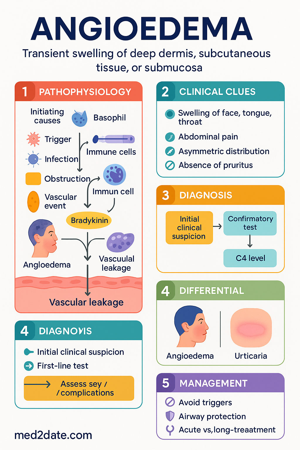
- Angioedema is transient, localised swelling of deep dermis, subcutaneous tissue, or submucosa; laryngeal involvement is a medical emergency with risk of fatal asphyxiation.
- Two principal pathophysiological pathways: histamine-mediated (allergic / mast-cell) and bradykinin-mediated (hereditary angioedema, ACE-inhibitor induced).
- Histamine-mediated angioedema typically responds to adrenaline, H₁-antihistamines, and corticosteroids; bradykinin-mediated angioedema does not — misdiagnosis delays effective treatment.
- Hereditary angioedema (HAE) is caused by C1-inhibitor deficiency (Type I ~85 %, Type II ~15 %); Type III (normal C1-INH) is rarer and often factor-XII mutation–related.
- ACE-inhibitor angioedema accounts for 20–40 % of emergency angioedema presentations in Australia; onset may occur months to years after commencing therapy.
- First-episode or diagnostic uncertainty requires C4 level as screening test — if low, proceed to C1-inhibitor quantitative and functional assays.
- Emergency airway management takes precedence; early anaesthetic/ENT review if any stridor, voice change, or tongue swelling.
- For histamine-mediated attacks: adrenaline 0.01 mg/kg IM (max 0.5 mg), IV dexchlorpheniramine or promethazine, hydrocortisone IV.
- For HAE acute attacks: C1-inhibitor concentrate (Berinert®) IV or icatibant (Firazyr®) SC — both available through special-access pathways in Australia.
- ACE inhibitors must be permanently discontinued after an angioedema episode; switch to ARB or alternative with monitoring.
- All patients with confirmed HAE should carry a personalised action plan and medical-alert identification; consider long-term prophylaxis with danazol, tranexamic acid, or subcutaneous C1-INH (Haegarda®).
- Aboriginal and Torres Strait Islander peoples may face delayed presentation due to geographic remoteness; ensure culturally safe follow-up and access to adrenaline autoinjectors.
Introduction & Australian Epidemiology
Angioedema is characterised by transient, localised subcutaneous or submucosal swelling resulting from increased vascular permeability in deep tissue layers. Unlike urticaria, which affects superficial dermis, angioedema involves deeper structures and presents with non-pitting, asymmetrical swelling — most commonly of the lips, tongue, periorbital region, and extremities. Involvement of the upper airway constitutes a life-threatening emergency that demands immediate recognition and intervention.
In Australia, angioedema accounts for an estimated 1 in 500 emergency department (ED) presentations annually, with hospital admission rates for angioedema rising by approximately 2–3 % per year over the past decade. The increasing prevalence is attributed in part to widespread ACE-inhibitor prescribing — ACE inhibitors are among the most commonly dispensed medications on the Pharmaceutical Benefits Scheme (PBS), and ACE-inhibitor angioedema now represents 20–40 % of all angioedema ED presentations nationally.
Hereditary angioedema (HAE) is rare, with an estimated prevalence of 1 in 50,000 Australians; however, significant diagnostic delay (averaging 8–10 years from first symptom to diagnosis) suggests under-recognition. The Australian HAE patient registry data indicate that patients experience a mean of 4–6 attacks per year before commencing prophylaxis, with laryngeal involvement reported in up to 50 % of patients over their lifetime.
This guideline covers the classification, pathophysiology, diagnosis, and emergency management of angioedema in the Australian clinical context, with attention to PBS-listed therapies, state-based hospital protocols, and equity considerations for underserved populations.
Types of Angioedema
Accurate classification is essential because histamine-mediated and bradykinin-mediated angioedema require fundamentally different treatments. The major types encountered in Australian practice are summarised below.
| Type | Mechanism | Common Triggers / Associations | Responds to Adrenaline / Antihistamines? |
|---|---|---|---|
| Allergic (histamine-mediated) | IgE-mediated mast-cell degranulation → histamine release | Foods (peanut, shellfish, egg), NSAIDs, latex, insect stings, antibiotics (penicillin) | Yes — first-line |
| Hereditary angioedema (HAE) Type I | Quantitative C1-inhibitor deficiency → unregulated kallikrein → excess bradykinin | Trauma, stress, dental procedures, OCP/oestrogens; often spontaneous | No |
| HAE Type II | Dysfunctional C1-inhibitor (normal or elevated levels, reduced function) | Same as Type I | No |
| HAE Type III (normal C1-INH) | Often factor-XII gain-of-function mutation; oestrogen-sensitive | Oestrogen-containing contraceptives, pregnancy | No |
| ACE-inhibitor angioedema | Bradykinin accumulation due to reduced degradation (ACE = kininase II) | Enalapril, ramipril, perindopril — may occur months–years after initiation | No |
| Acquired C1-inhibitor deficiency | Autoantibody consumption of C1-INH; associated with lymphoproliferative disorders | B-cell lymphoma, monoclonal gammopathy | No |
| Idiopathic | Unknown; may be histamine-mediated or bradykinin-mediated | Diagnosis of exclusion | Variable |
Pathophysiology
Histamine-Mediated (Allergic) Pathway
Allergen cross-links surface-bound IgE on mast cells and basophils, triggering degranulation and release of histamine, tryptase, prostaglandins, and leukotrienes. Histamine acts on H₁-receptors on vascular endothelial cells, causing endothelial cell contraction, intercellular gap formation, and plasma extravasation into the deep dermis and submucosa. The result is non-pitting oedema that is typically pruritic, often accompanied by urticaria, and resolves within 24–72 hours.
Bradykinin-Mediated Pathway
Bradykinin is a potent vasoactive nonapeptide generated by the action of kallikrein on high-molecular-weight kininogen (HMWK). C1-inhibitor (C1-INH) is the primary regulatory serine protease inhibitor (serpin) of this pathway — it inhibits activated factor XII (Hageman factor), plasma kallikrein, and C1s/C1r in the classical complement pathway.
In HAE Type I (~85 % of cases), reduced synthesis of C1-INH protein leads to uninhibited kallikrein activity and excess bradykinin production. In Type II (~15 %), C1-INH protein levels are normal or elevated but the molecule is dysfunctional. In both types, C4 is chronically low because of unregulated classical-pathway consumption.
ACE-inhibitor angioedema occurs because angiotensin-converting enzyme (ACE) is identical to kininase II — the primary enzyme responsible for bradykinin degradation. Inhibition of ACE raises local bradykinin concentrations, predisposing to angioedema. Individual susceptibility likely depends on polymorphisms in aminopeptidase P and neprilysin (alternative bradykinin-degradation pathways).
Clinical Features & Diagnosis
Clinical Presentation
Angioedema presents with recurrent episodes of localised, non-pitting oedema affecting one or more of the following sites:
- Face: Lips, periorbital region, cheeks — the most common site overall
- Upper airway: Tongue, uvula, soft palate, larynx — potentially fatal; 25–50 % of HAE patients experience at least one laryngeal attack
- Extremities: Hands, feet — can be debilitating and interfere with daily function
- Gastrointestinal tract: Colicky abdominal pain, nausea, vomiting, diarrhoea — due to bowel-wall oedema; may mimic an acute abdomen
- Urogenital: Genital swelling (more common in HAE)
Differentiating Histamine-Mediated from Bradykinin-Mediated Angioedema
| Feature | Histamine-Mediated | Bradykinin-Mediated (HAE / ACE-I) |
|---|---|---|
| Urticaria | Usually present (≥ 90 %) | Absent |
| Pruritus | Prominent | Usually absent; may have tingling / tightness |
| Prodrome | None or sudden onset | Tingling, erythema marginatum (serpiginous, non-pruritic rash) in ~40 % of HAE |
| Onset to peak | Minutes to 1–2 hours | Gradual over 12–36 hours; untreated lasts 2–5 days |
| Response to adrenaline | Rapid improvement | Poor or absent |
| Family history | Atopy | Autosomal dominant (50 %); 25 % are de-novo mutations |
| Medications | NSAIDs, antibiotics | ACE inhibitors, oestrogens |
Diagnostic Approach
The diagnostic algorithm depends on clinical context:
Investigations with Australian Availability
Emergency Management
Immediate Resuscitation (All Angioedema Types)
Histamine-Mediated (Allergic) Angioedema — Acute Treatment
Bradykinin-Mediated Angioedema (HAE) — Acute Treatment
Standard allergy therapies (adrenaline, antihistamines, corticosteroids) are not effective for bradykinin-mediated attacks. Targeted therapies are required and should be administered as early as possible.
ACE-Inhibitor Angioedema — Management
If the attack is bradykinin-mediated and targeted HAE therapies are available, consider icatibant or C1-INH concentrate under specialist guidance. Evidence is emerging but not yet definitive. Icatibant has shown benefit in small RCTs for ACE-inhibitor angioedema.
For ACE-inhibitor angioedema with airway compromise that is not responding to standard measures, discuss with the on-call immunologist or contact the Australian HAE Helpline (through HAE Australasia) for advice on emergency access to C1-INH concentrate.
Observation & Disposition
Long-Term Prophylaxis for HAE
Long-term prophylaxis (LTP) is indicated for patients with ≥ 1 attack per month, significant disease burden, laryngeal attack history, or limited access to on-demand therapy. Options available in Australia:
Special Populations
Aboriginal and Torres Strait Islander Health Considerations
📚 References
- 1. Maurer M, Magerl M, Ansotegui I, et al. The international WAO/EAACI guideline for the management of hereditary angioedema — the 2021 revision and update. World Allergy Organ J. 2022;15(1):100627.
- 2. Betschel S, Badiou J, Binkley K, et al. The International/Canadian Hereditary Angioedema Guideline. Allergy Asthma Clin Immunol. 2019;15:72.
- 3. Australasian Society of Clinical Immunology and Allergy (ASCIA). ASCIA guidelines for prevention of anaphylaxis in schools, preschools and childcare. Sydney: ASCIA; 2023.
- 4. Cicardi M, Aberer W, Banerji A, et al. Classification, diagnosis, and approach to treatment for angioedema: consensus report from the Hereditary Angioedema International Working Group. Allergy. 2014;69(5):602–616.
- 5. Bernstein JA, Cremonesi A, Hoffmann TK, Hollingsworth J. Angioedema in the emergency department: a practical guide to differential diagnosis and management. Int J Emerg Med. 2017;10(1):15.
- 6. Banerji A, Clark S, Bland K, et al. ACE-inhibitor angioedema in the emergency department: clinical characteristics, management, and outcomes. J Allergy Clin Immunol Pract. 2020;8(10):3385–3392.
- 7. Pharmaceutical Benefits Scheme (PBS). Australian Government Department of Health. Available at: https://www.pbs.gov.au. Accessed June 2025.
- 8. Australian Commission on Safety and Quality in Health Care (ACSQHC). National Safety and Quality Health Service Standards. 2nd ed. Sydney: ACSQHC; 2021.
- 9. Australian Institute of Health and Welfare (AIHW). Allergic disease in Australia. Cat. no. ACM 35. Canberra: AIHW; 2023.
- 10. HAE Australasia. Living with hereditary angioedema in Australia and New Zealand. Patient registry report. Sydney: HAE Australasia; 2023.
- 11. Frank MM, Gelfand JA, Atkinson JP. Hereditary angioedema: the clinical syndrome and its management. Ann Intern Med. 1976;84(5):580–593.
- 12. RHDAustralia (Northern Territory Department of Health and Menzies School of Health Research). The Australian guidelines for prevention, diagnosis and management of acute rheumatic fever and rheumatic heart disease. 3rd ed. Darwin: RHDAustralia; 2020. [Referenced for ATSI health frameworks applicable across clinical guidelines.]
- 13. Sinert R, Levy P, Bernstein JA, et al. Randomized trial of icatibant for ACE-inhibitor-induced angioedema in the emergency department. Ann Emerg Med. 2017;69(4):389–397.
- 14. Lumry WR, Li HH, Levy RJ, et al. Randomized placebo-controlled trial of the bradykinin B₂ receptor antagonist icatibant for the treatment of acute attacks of hereditary angioedema: the FAST-3 trial. Ann Allergy Asthma Immunol. 2011;107(6):529–537.
- 15. The Australasian Society of Clinical Immunology and Allergy (ASCIA). Position statement — prevention of anaphylaxis. Sydney: ASCIA; 2024.
📚 References
- 1. Australian Institute of Health and Welfare (AIHW). Autoimmune disease in Australia. Cat. no. PHE 312. Canberra: AIHW; 2023.
- 2. Fraenkel L, Bathon JM, England BR, et al. 2021 American College of Rheumatology guideline for the treatment of rheumatoid arthritis. Arthritis Care Res. 2021;73(7):924–939.
- 3. Fanouriakis A, Kostopoulou M, Alber K, et al. 2019 update of the EULAR recommendations for the management of systemic lupus erythematosus. Ann Rheum Dis. 2019;78(6):736–745.
- 4. Chung SA, Langford CA, Maz M, et al. 2021 American College of Rheumatology/Vasculitis Foundation guideline for the management of antineutrophil cytoplasmic antibody-associated vasculitis. Arthritis Care Res. 2021;73(11):1583–1599.
- 5. Smolen JS, Landewé RBM, Bijlsma JWJ, et al. EULAR recommendations for the management of rheumatoid arthritis with synthetic and biological disease-modifying antirheumatic drugs: 2022 update. Ann Rheum Dis. 2023;82(1):3–18.
- 6. Australian Technical Advisory Group on Immunisation (ATAGI). Australian Immunisation Handbook. Australian Government Department of Health; 2024. Available from: immunisationhandbook.health.gov.au.
- 7. Rheumatic Heart Disease Australia (RHDAustralia). The 2020 Australian guideline for prevention, diagnosis, and management of acute rheumatic fever and rheumatic heart disease. 3rd ed. Darwin: Menzies School of Health Research; 2020.
- 8. Pharmaceutical Benefits Scheme (PBS). PBS Schedule. Australian Government Department of Health. Available from: pbs.gov.au. Accessed 2024.
- 9. Agarwal S, Cunnington J, Nossent J. Autoimmune disease in Indigenous Australians: a systematic review. Int J Rheum Dis. 2021;24(12):1487–1498.
- 10. Pisetsky DS. Antinuclear antibody testing — misunderstood or misused? Clin Immunol. 2023;255:109717.
- 11. Bertsias GK, Tektonidou M, Amoura Z, et al. Joint European League Against Rheumatism and European Renal Association–European Dialysis and Transplant Association (EULAR/ERA-EDTA) recommendations for the management of adult and paediatric lupus nephritis. Ann Rheum Dis. 2012;71(11):1771–1782.
- 12. Ledingham J, Deighton C; British Society for Rheumatology Standards, Audit and Guidelines Working Group. Update on the British Society for Rheumatology guidelines for prescribing TNFα blockers in adults with rheumatoid arthritis. Rheumatology. 2005;44(2):155–158.
- 13. National Health and Medical Research Council (NHMRC). National statement on ethical conduct in human research. Canberra: NHMRC; 2023 (updated).
📚 References
- 1. Australian Institute of Health and Welfare (AIHW). Autoimmune disease in Australia. Cat. no. PHE 312. Canberra: AIHW; 2023.
- 2. Fraenkel L, Bathon JM, England BR, et al. 2021 American College of Rheumatology guideline for the treatment of rheumatoid arthritis. Arthritis Care Res. 2021;73(7):924–939.
- 3. Fanouriakis A, Kostopoulou M, Alber K, et al. 2019 update of the EULAR recommendations for the management of systemic lupus erythematosus. Ann Rheum Dis. 2019;78(6):736–745.
- 4. Chung SA, Langford CA, Maz M, et al. 2021 American College of Rheumatology/Vasculitis Foundation guideline for the management of antineutrophil cytoplasmic antibody-associated vasculitis. Arthritis Care Res. 2021;73(11):1583–1599.
- 5. Smolen JS, Landewé RBM, Bijlsma JWJ, et al. EULAR recommendations for the management of rheumatoid arthritis with synthetic and biological disease-modifying antirheumatic drugs: 2022 update. Ann Rheum Dis. 2023;82(1):3–18.
- 6. Australian Technical Advisory Group on Immunisation (ATAGI). Australian Immunisation Handbook. Australian Government Department of Health; 2024. Available from: immunisationhandbook.health.gov.au.
- 7. Rheumatic Heart Disease Australia (RHDAustralia). The 2020 Australian guideline for prevention, diagnosis, and management of acute rheumatic fever and rheumatic heart disease. 3rd ed. Darwin: Menzies School of Health Research; 2020.
- 8. Pharmaceutical Benefits Scheme (PBS). PBS Schedule. Australian Government Department of Health. Available from: pbs.gov.au. Accessed 2024.
- 9. Agarwal S, Cunnington J, Nossent J. Autoimmune disease in Indigenous Australians: a systematic review. Int J Rheum Dis. 2021;24(12):1487–1498.
- 10. Pisetsky DS. Antinuclear antibody testing — misunderstood or misused? Clin Immunol. 2023;255:109717.
- 11. Bertsias GK, Tektonidou M, Amoura Z, et al. Joint European League Against Rheumatism and European Renal Association–European Dialysis and Transplant Association (EULAR/ERA-EDTA) recommendations for the management of adult and paediatric lupus nephritis. Ann Rheum Dis. 2012;71(11):1771–1782.
- 12. Ledingham J, Deighton C; British Society for Rheumatology Standards, Audit and Guidelines Working Group. Update on the British Society for Rheumatology guidelines for prescribing TNFα blockers in adults with rheumatoid arthritis. Rheumatology. 2005;44(2):155–158.
- 13. National Health and Medical Research Council (NHMRC). National statement on ethical conduct in human research. Canberra: NHMRC; 2023 (updated).