Here , we are going to discuss a diagnostic approach to Systemic Lupus Erythematosus involving clinical picture , Laboratory investigations and summary of diagnostic criteria .


Clinical picture
• Male: female ratio is 1 : 9
• The presentation and course are highly variable :
1- Fever of unknown origin.
2- Musculoskeletal manifestations :

- Joint
involvement is mainly arthralgia with mild morning stiffness.
- The
arthropathy is bilateral and symmetrical. The small joints are
usually affected mimic rheumatoid disease.
- It is non
deforming but tendosynovitis may lead to deformity (Jaccoud's arthropathy), it
is due to tendon or ligament laxity.
- A vascular
necrosis of the hip may occur with steroid therapy.
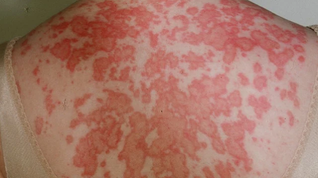
3- The skin
-
Butterfly rash: fixed erythema (flat or raised) on the cheeks of the face
and across the bridge of the nose, occurs in a photosensitive distribution that
spares the naso-labial folds.
atrophic
scarring may occur. It may lead to scarring alopecia if present on the scalp.
-
Photosensitivity: skin rash as a result of unusual reaction to sun light.
-
Purpuric lesion due to thrombocytopenia or vasculitis.
- Leg
Ulcers .
Vasculitic lesions :
• Nail bed and
finger bulb infarcts .
• Purpuric
rash with elevated edge.
- Raynaud's phenomenon.
- Urticaria. - Alopecia.
- Panniculitis (Lupus profundus)
- Lichen plannus like.
- Livedo reticularis.
4- The Eye
- Retinal vasculitis can cause infarcts, cytoid
bodies which appear as hard exudates
- Episcleritis, conjunctivitis or optic
neuritis may occur.
- Kerataconjunctivitis sicca with Sjogren's
syndrome.
5- The heart
- Pericarditis and pericardial effusion.
- Myocarditis with heart failure.
- Libman sacks endocarditis (affecting mitral
or aortic valves causing regurge), It is a sterile endocarditis.
- Blood pressure is increased with renal
hypertension.
- Coronary heart disease (accelerated
atherosclerosis).
6- The Kidney
Lupus nephritis (WHO classification)
• Type I Minimal
pathology (Normal glomeruli)
• Type II Mesangial
widening with or without hypercellularity.
• Type III Focal
proliferative G.N.
• Type IV Diffuse
proliferative G.N.
• Type V Membranous
G.N.
• Type VI Advancing sclerosing G.N.
7- GIT
- Mesenteric vasculitis with acute abdomen.
Liver involvement is unusual, pancreatitis
is uncommon.
- Nausea, vomiting and diarrhea can occur with
an SLE flare.
8- The Lung
- Pleurisy and pleural effusion
- Interstitial pulmonary fibrosis.
- Shrinking lung syndrome with elevation of the
diaphragm due to recurrent pulmonary
infarction.
- Pulmonary hypertension with antiphospholipid
syndrome.
- Adult respiratory distress syndrome.
9- Neuro
psychiatric manifestations
- Psychosis, depression, cognitive dysfunction
(difficulties with memory and resoning).
- Lymphocytic
meningitis, transverse myelitis.
- Chorea.
- Cerebral vasculitis leading to cerbrovascular
stroke.
- PolyNeuropathy .
- Lupus headache. - Seizures .
Psychosis due to lupus must be differentiated from
steroid induced psychosis which occurs in the first weeks of steroid therapy at
doses of 2 40 mg of prednisone or equivalent, it resolves over several days
after steroids are decreased or stopped .
10-Blood
- Autoimmune thrombocytopenia and haemolytic
anaemia .
- Lymphopenia (guide to disease activity).
- Antiphospholipid $ leading to thrombo-embolism .
11- Polyserositis affecting:
- Pleura. -
Pericardium. - Peritoneum.

Diagnostic Criteria of Systemic Lupus Erythematosus
1. Butterfly rash 50% : Fixed erythema- flat OR -
raised
2. Discoid rash 20% : Erythymatous raised patches + scales
3. Photosensitivity 70% : Rash on exposure to sun
light
4. Oral Ulcers 40% : Painless, it may be nasopharyngeaL
5- ArthroPathy 95% : Involving 2 or more peripheral joints.
6- Serositis. : Pleuritis, pericarditis
7- Renal (50%have
clinical nephritis) persistent protinuria >
0.5 grn/24h (30-50%)
Casts and RBCs .
8. Neurologic disorders
Seizures or psychosis in the absence of offending drugs
or known metabolic disorders .
- Hematological disorders
- Leukopenia < 4000 / mm
- Lymphopenia < 1500 / mm
- Thrombocytopenia < 100000 /
mm
- Hemolytic anemia
10 . Immunologic disorders
- Anti DNA
- Anti-sm antibody
- Anti-phospholipid antibody
- Abnormal titre of ANA .
To diagnose patients with SLE, 4 or more criteria
must be present serillay or simultaneously or have occurred in the
past .
LAB investigations
1- Blood
•
Anemia of chronic disease (normocytic normochromic).
•
Autoimmune hemolytic anaemia (positive coomb's test).
•
Leucopenia, lymphopenia with activity, thrombocytopenia.
2- ESR is High with activity of the disease.
3- Immunological tests
a- C-reactive protein is low but increases with superimposed infection.
b- Hyper gammaglobulinemia usually IgG and IgM
(polyclonal).
c- Low C3
& C4 as they are consumed during disease activity.
d- ANA + ve (it is positive in almost all
cases, 95%), patients with negative ANA are unlikely to have SLE.
e- Anti DNA is the most specific. It is positive in
about 60% of cases, it may reflect disease activity .
f- Rheumatoid factor is positive in 30 % of cases.
g- Anti Ro, La antibodies (They are asked if ANA is
negative, especially anti-Ro).
Anti Ro is the causal antibody for neonatal lupus and
congenital
heart block.
h- Anti-sm Ab (specific for SLE).
4- Kidney function tests and urine analysis for
protein, RBCS and casts to detect renal involvement.
Q : Symptoms and signs suggesting active SLE?
• Weight loss, fever, arthritis, seizures, hair loss,
anaemia, haematuria, rashes, mouth sores and oliguria.
Q : Laboratory
diagnosis of disease activity?
• -Low C3 , C4
• +ve Anti-DNA (high titre)
• Disease activity (see above)
• Infection (+ ve C- reactive protein)
• Steroid therapy.
- PNL high -Eosinophils low - Lymphocytes low .
• Infection:
- Toxic granulations within WBCs, presence of staff
cells and positive CRP.
Sequence of investigations to diagnose SLE

Subsets of Lopus
A- Idiopathic
• Systemic lupus.
• Chronic discoid lupus (CDLE) is a benign variant of
the disease in which skin involvement is often the only feature, systemic manifestations
may occur with time (5%)
ANA is
positive in 30%.
• Subacute cutaneous lupus, -ve ANA, +ve anti Ro, anti La, organ involvement is rare.
• Late onset after 50 years age.
.. Neonatal Lupus with positive Ab to Ro. and La.
B- Drug induced (see before)
C- Overlap
syndrome