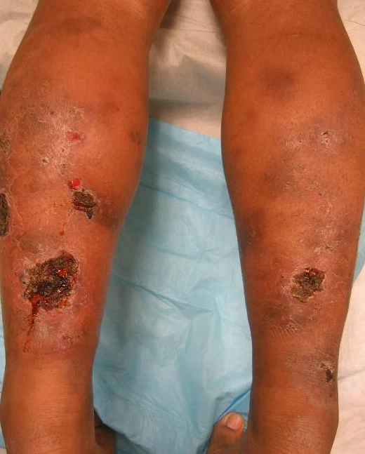
Polyarteritis nodosa can occur at any age but usually occur in the 40s and 50s with male : female ratio 2 : 1, it is a necrotizing arteritis, with transmural inflammation affecting medium sized arteries.
Etiology
1- Circulating immune complex with low C3, C4
2- Hepatitis HBsAg +ve in 25 % (serum sickness like syndrome), also there is an association with HCV.
3- Other viruses are implicated e.g. hepatitis A, CMV, parvovirus.
1- Kidney (The most common involved organ)
• Affection of arcuate arteries with narrowing ---7 multiple renal infarcts 7 renal hypertension and renal
impairement.
• Rapidly progressive glomerulonephr:itis may occur.
• Spontaneous rupture of aneurysms can result in retroperitoneal hemorrhage or perinephric hematoma.
2- Lung (involvement is rare)
• Pleurisy, lung infiltrates may occur.
3- Skin lesion
• Palpable purpura, infarcts.
• Livedo reticularis, urticaria. • Ulcers.
4- CVS
• Coronary heart disease (angina pectoris or infarction).
• Pericarditis may occur.
5- Nervous system.
• Stroke.
• Mononeuritis multiplex (arteritis in vasa nervorum) .
6- Musculoskeletal:
• Arthralgia, myalgia.
7- Abdomen
• Mesenteric occlusion? acute abdomen.
• Intestinal bleeding.
• Pancreatitis.
Pathology
Lab. Investigations
• High ESR and + ve CRP
• Low Hb , platelets increase .
• ANA -ve , if it is positive it is mostly lupus vasculitis !?
• Rh.F -ve , if it is positive it is mostly RA complicated with vasculitis.
• HB s Ag +ve in 25 %
• Low Cs , C4 (Hypocomplementemia), ANCA is rarely positive.
• Tissue biopsy from the kidney, muscle or sural nerve.
• Eosinophilia.
• Angiography of renal, hepatic or mesenteric arteries showing multiple micro aneurysms.
Treatment
• The recommended initial therapy is prednisone 1-2 mg / kg / d and cyclophosphamide 2- mg. / kg / d, gradual tapering with
improvement, then maintenance therapy to maintain remission.
• Steroid plus interferon if there is positive hepatitis B or C.
Etiology
1- Circulating immune complex with low C3, C4
2- Hepatitis HBsAg +ve in 25 % (serum sickness like syndrome), also there is an association with HCV.
3- Other viruses are implicated e.g. hepatitis A, CMV, parvovirus.
How to diagnose Polyarteritis nodosa ?
Clinical picture (Vascular occlusion)1- Kidney (The most common involved organ)
• Affection of arcuate arteries with narrowing ---7 multiple renal infarcts 7 renal hypertension and renal
impairement.
• Rapidly progressive glomerulonephr:itis may occur.
• Spontaneous rupture of aneurysms can result in retroperitoneal hemorrhage or perinephric hematoma.
2- Lung (involvement is rare)
• Pleurisy, lung infiltrates may occur.
3- Skin lesion
• Palpable purpura, infarcts.
• Livedo reticularis, urticaria. • Ulcers.
4- CVS
• Coronary heart disease (angina pectoris or infarction).
• Pericarditis may occur.
5- Nervous system.
• Stroke.
• Mononeuritis multiplex (arteritis in vasa nervorum) .
6- Musculoskeletal:
• Arthralgia, myalgia.
7- Abdomen
• Mesenteric occlusion? acute abdomen.
• Intestinal bleeding.
• Pancreatitis.
Pathology
Lab. Investigations
• High ESR and + ve CRP
• Low Hb , platelets increase .
• ANA -ve , if it is positive it is mostly lupus vasculitis !?
• Rh.F -ve , if it is positive it is mostly RA complicated with vasculitis.
• HB s Ag +ve in 25 %
• Low Cs , C4 (Hypocomplementemia), ANCA is rarely positive.
• Tissue biopsy from the kidney, muscle or sural nerve.
• Eosinophilia.
• Angiography of renal, hepatic or mesenteric arteries showing multiple micro aneurysms.
Treatment
• The recommended initial therapy is prednisone 1-2 mg / kg / d and cyclophosphamide 2- mg. / kg / d, gradual tapering with
improvement, then maintenance therapy to maintain remission.
• Steroid plus interferon if there is positive hepatitis B or C.

