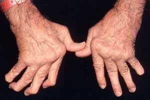
• Rheumatoid arthritis (RA) is the most common inflammatory arthritis and hence an important cause of potentially preventable disability.
Definition
• It is a chronic systemic disease leading to symmetrical inflammatory polyarthritis affecting mainly peripheral joints, with progressive joint
damage, also it is associated with extra-articular manifestations.
- Unknown (triggered by T lymphocyte activation)
- Possible factors I?:
• Autoimmune (infiltration of synovial membrane with plasma cells and lymphocytes).
• HLA association e.g. in HLA-DR4.
• Infection, bacterial or slow virus infections have been implicated (no definite evidence).
• Smoking also is a risk factor for RA and for positivity of rheumatoid factor in non RA subjects!?
• Production of autoantibodies or rheumatoid factors (lg M)in the blood which react with Fc altered portion of Ig Gwith activation of the complement, causing immune complex in synovial membranes with release of intlarnrnatory.rnediators, cytokines e.g TNF,. IL1, IL6.
Rheumatoid factors are positive (sero positive cases) in 80% of cases of rheumatoid disease.
The triggering, antigen remain unclear , it is suggested that the glycosylation pattern of immunoglobulins may be abnormal in RA rendering them potentially antigenic.
• T lymphocyte activation and macrophages in genetically predisposed persons e.g. HLA-DR4.
• The presence of activated· T cells and macrophages and production of rheumatoid factor autoantibodies in RA suggests that immune dysregulation plays a major role in pathogenesis.
Pathology
The earliest change is swelling and congestion of the synovial membrane and the underlying connective tissue .
• Synovial membrane is infiltrated with lymphocytes, plasma cells and macrophages.
Effusion of synovial fluid into the joint space takes place during active phases of the disease.
• The inflamed synovial membrane becomes thick oedematous and proliferating forming villi filling the joint space.
Later an inflammatory granulation tissue is formed ( Pannus ) spreads over and under the articular cartilage which is progressively eroded and destroyed.
• By time pannus will be organised ---> fibrous tissue ---> ankylosis of joint.
b- Capsule ---> Thickened with fibrosis.
c- Juxta articular bone ---> osteoporosis.
Rheumatoid nodules (subcutaneous)
• They are granulomatous lesions that occur in approximately 20 % of patients (almost exclusively in seropositive patients).
• Present at sites of friction and over pressure points.
• Tendon sheath.
• Extensor of the forearm and sacrum.
It is a central zone of fibrinoid material and surrounded by a palisade of proliferating mononuclear cells ..
Important links to visit :Definition
• It is a chronic systemic disease leading to symmetrical inflammatory polyarthritis affecting mainly peripheral joints, with progressive joint
damage, also it is associated with extra-articular manifestations.
What are the causes and triggers of Rheumatoid arthritis ?
Aetiology- Unknown (triggered by T lymphocyte activation)
- Possible factors I?:
• Autoimmune (infiltration of synovial membrane with plasma cells and lymphocytes).
• HLA association e.g. in HLA-DR4.
• Infection, bacterial or slow virus infections have been implicated (no definite evidence).
• Smoking also is a risk factor for RA and for positivity of rheumatoid factor in non RA subjects!?
How Rheumatoid arthritis develops ?
Pathogenesis• Production of autoantibodies or rheumatoid factors (lg M)in the blood which react with Fc altered portion of Ig Gwith activation of the complement, causing immune complex in synovial membranes with release of intlarnrnatory.rnediators, cytokines e.g TNF,. IL1, IL6.
Rheumatoid factors are positive (sero positive cases) in 80% of cases of rheumatoid disease.
The triggering, antigen remain unclear , it is suggested that the glycosylation pattern of immunoglobulins may be abnormal in RA rendering them potentially antigenic.
• T lymphocyte activation and macrophages in genetically predisposed persons e.g. HLA-DR4.
• The presence of activated· T cells and macrophages and production of rheumatoid factor autoantibodies in RA suggests that immune dysregulation plays a major role in pathogenesis.
Pathology
The earliest change is swelling and congestion of the synovial membrane and the underlying connective tissue .
• Synovial membrane is infiltrated with lymphocytes, plasma cells and macrophages.
Effusion of synovial fluid into the joint space takes place during active phases of the disease.
• The inflamed synovial membrane becomes thick oedematous and proliferating forming villi filling the joint space.
Later an inflammatory granulation tissue is formed ( Pannus ) spreads over and under the articular cartilage which is progressively eroded and destroyed.
• By time pannus will be organised ---> fibrous tissue ---> ankylosis of joint.
b- Capsule ---> Thickened with fibrosis.
c- Juxta articular bone ---> osteoporosis.
Rheumatoid nodules (subcutaneous)
• They are granulomatous lesions that occur in approximately 20 % of patients (almost exclusively in seropositive patients).
• Present at sites of friction and over pressure points.
• Tendon sheath.
• Extensor of the forearm and sacrum.
It is a central zone of fibrinoid material and surrounded by a palisade of proliferating mononuclear cells ..
- Treatment of Rheumatoid arthritis
- Investigations and monitoring of Rheumatoid Arthritis
- Diagnostic Criteria of rheumatoid arthritis and DD from rheumatic fever
- Extra-articular manifestations of Rheumatoid arthritis
- Clinical picture of Rheumatoid arthritis , symptoms and signs

