📋 Key Information Summary
- Melanoma is the most common cancer in Australians aged 15–39 years, with the highest incidence worldwide.
- Australia recorded ~18,200 new melanoma cases in 2024; age-standardised rate ≈ 49 per 100,000.
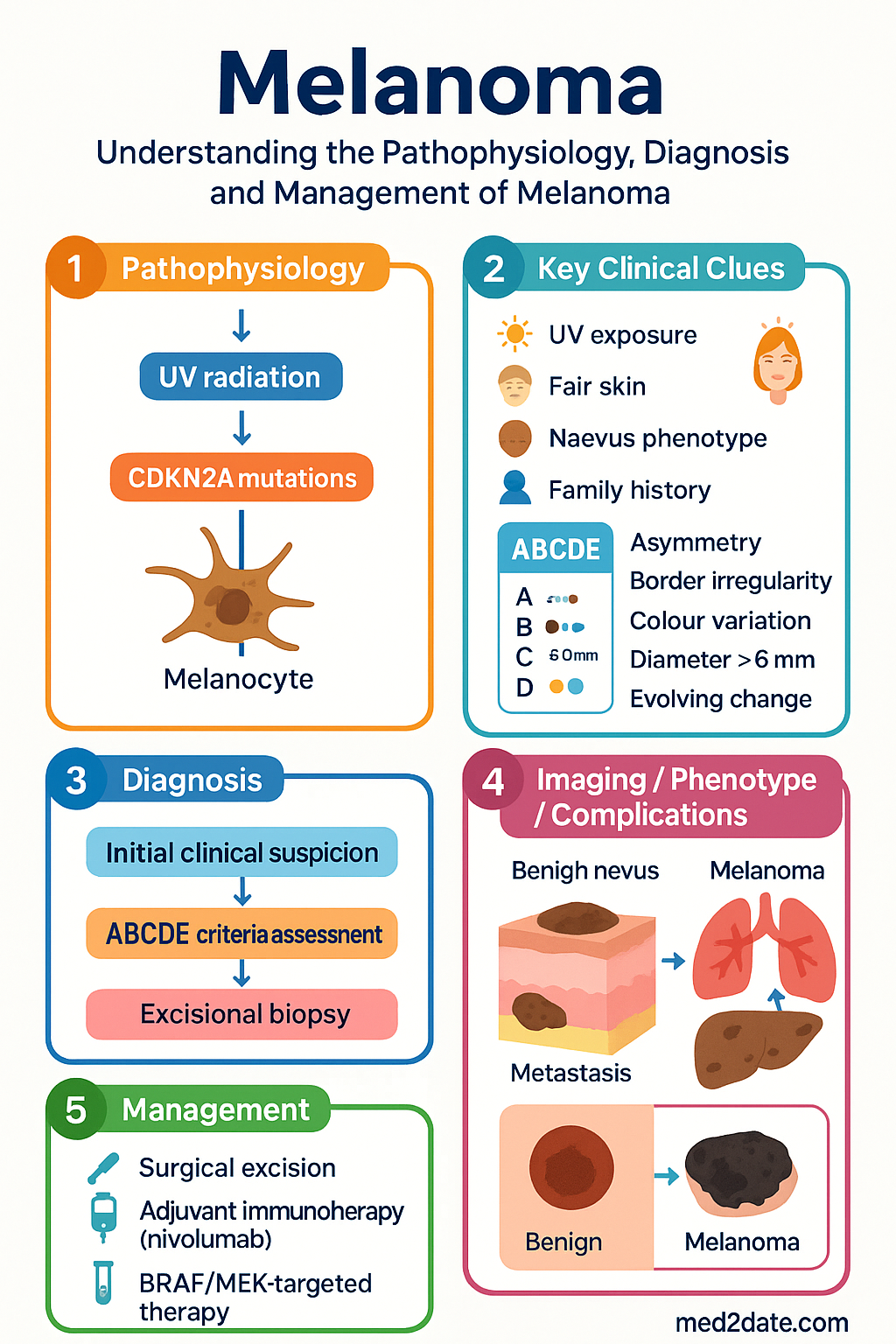
- Major risk factors include UV radiation exposure, fair skin, naevus phenotype, family history, and germline CDKN2A mutations.
- The ABCDE mnemonic (Asymmetry, Border irregularity, Colour variation, Diameter >6 mm, Evolving change) remains the clinical standard for early detection.
- TNM staging (8th AJCC edition) guides prognosis; sentinel lymph node biopsy (SLNB) is standard of care for primary tumours >1 mm Breslow thickness.
- Stage I–IIC (localised) disease: surgical excision with appropriate margins (0.5–2 cm) is curative in the majority.
- Stage III (regional) disease: adjuvant immunotherapy (nivolumab) or BRAF/MEK-targeted therapy (dabrafenib + trametinib) significantly reduce recurrence risk.
- Stage IV (metastatic) disease: combination immunotherapy (nivolumab + ipilimumab) or BRAF/MEK inhibition if BRAF V600-mutated.
- Immunotherapy-related adverse events (irAEs) require prompt recognition; endocrine, hepatic, and colonic toxicities are most common.
- Aboriginal and Torres Strait Islander Australians have higher late-stage presentation rates and reduced access to specialist melanoma services, particularly in remote settings.
- All patients require lifelong UV protection counselling, regular skin surveillance, and structured follow-up schedules.
- Key PBS-listed melanoma agents: pembrolizumab, nivolumab, ipilimumab, dabrafenib, trametinib, vemurafenib, and encorafenib — all Authority Required or PBS-listed under Section 100 for specified indications.
Introduction & Australian Epidemiology
Melanoma arises from the malignant transformation of melanocytes and is distinguished by its capacity for early lymphatic and haematogenous dissemination. In Australia, melanoma represents the third most commonly diagnosed cancer overall and remains a leading cause of cancer death in young and middle-aged adults. When detected at an early stage, surgical excision alone achieves cure rates exceeding 95%; however, metastatic disease historically carried a median survival of less than one year prior to the advent of immunotherapy and molecular-targeted agents.
Australia's melanoma burden is unparalleled globally, driven by a predominantly fair-skinned population, high ambient UV index, and outdoor lifestyle. National data from the Australian Institute of Health and Welfare (AIHW) demonstrate that melanoma incidence has continued to rise among both sexes, while mortality rates have plateaued owing to improved surveillance and therapeutic advances.
| Parameter | Value |
|---|---|
| New cases (2024 est.) | ~18,200 |
| Deaths (2024 est.) | ~1,300 |
| 5-year relative survival (all stages) | 92% (men), 95% (women) |
| Age-standardised incidence | ~49 per 100,000 |
| Lifetime risk (men) | 1 in 14 |
| Lifetime risk (women) | 1 in 23 |
| Median age at diagnosis | 64 years |
| Most common site (men / women) | Trunk / Lower limb |
Epidemiology & Risk Factors
Environmental Risk Factors
- Ultraviolet radiation (UVR): The dominant environmental risk factor. Intermittent high-intensity sun exposure and sunburn in childhood/adolescence confer the greatest risk. Occupational and cumulative UVR exposure is also relevant.
- Geographic variation: Residents of Queensland face the highest state-level incidence (ASR ~72 per 100,000 in males) compared to Tasmania (~35 per 100,000).
- Solarium exposure: Artificial UV exposure before age 35 increases melanoma risk by 59%. Solariums have been banned in all Australian states and territories since 2016.
- Latitude and altitude: Higher UV-B flux at lower latitudes and higher altitudes increases cumulative dose.
Host Risk Factors
- Skin phenotype: Fitzpatrick skin types I and II (always burn, rarely tan) carry the highest risk.
- Naevus phenotype: Greater than 50 common naevi, or presence of atypical (dysplastic) naevi, significantly elevate risk.
- Freckling: Extensive facial or body freckling is an independent risk marker.
- Family history: First-degree relative with melanoma confers a two- to three-fold increased risk. Familial melanoma syndromes (CDKN2A, CDK4, BAP1 germline mutations) account for approximately 5–10% of cases.
- Personal history: Prior melanoma or non-melanoma skin cancer significantly increases future melanoma risk.
- Immunosuppression: Organ transplant recipients and patients on chronic immunosuppressive therapy have a 2–3-fold elevated risk with more aggressive biology.
Other Associations
- Congenital melanocytic naevi: Giant congenital naevi (>20 cm) carry a 5–10% lifetime risk of malignant transformation.
- Xeroderma pigmentosum: Autosomal recessive DNA repair defect — melanoma risk increased >2000-fold.
- Non-cutaneous melanoma: Mucosal and uveal melanoma have distinct risk profiles unrelated to UV exposure.
Pathology & ABCDE Classification
Histological Subtypes
Accurate histopathological subtyping is critical for prognosis, staging, and treatment planning. The four principal subtypes of cutaneous melanoma are:
| Subtype | Frequency | Typical Location | Key Features | Prognosis |
|---|---|---|---|---|
| Superficial spreading melanoma (SSM) | ~60–70% | Trunk, limbs | Flat, irregular, variable pigment; radial growth phase | Good if thin |
| Nodular melanoma (NM) | ~15–30% | Trunk, head & neck | Rapid vertical growth; may be amelanotic | Worse (thicker at Dx) |
| Lentigo maligna melanoma (LMM) | ~5–10% | Sun-damaged face | Large, flat, slow-growing; elderly patients | Good (slow invasion) |
| Acral lentiginous melanoma (ALM) | ~5% | Soles, palms, subungual | Higher proportion in darker skin types; less UV-related | Often diagnosed late |
The ABCDE Mnemonic
The ABCDE criteria provide a structured framework for clinical assessment of pigmented lesions:
Histopathological Reporting (Structured)
Every melanoma histopathology report in Australia should include the following data elements as recommended by the Royal College of Pathologists of Australasia (RCPA):
- Breslow thickness (mm) — measured from the granular layer or ulcer base
- Ulceration status (present or absent)
- Mitotic rate (per mm²) — particularly important for thin melanomas
- Clark level of invasion (I–V)
- Margins — peripheral and deep excision clearance
- Lymphovascular invasion
- Microsatellitosis
- Tumour-infiltrating lymphocytes (TILs) — brisk, non-brisk, absent
- Regression (present or absent, percentage)
- Neurotropism
Staging & Sentinel Lymph Node Biopsy
AJCC 8th Edition TNM Staging
The American Joint Committee on Cancer (AJCC) 8th edition TNM system, adopted universally in Australia, remains the current standard for melanoma staging. Staging incorporates tumour thickness, ulceration, mitotic rate, nodal status, and distant metastases.
T Stage (Primary Tumour)
| T Stage | Breslow Thickness | Ulceration |
|---|---|---|
| Tis | Melanoma in situ | — |
| T1a | ≤0.8 mm | No ulceration, mitosis <1/mm² |
| T1b | ≤0.8 mm with ulceration/↑ mitotic rate OR 0.8–1.0 mm | Variable |
| T2a | 1.0–2.0 mm | No ulceration |
| T2b | 1.0–2.0 mm | Ulcerated |
| T3a | 2.0–4.0 mm | No ulceration |
| T3b | 2.0–4.0 mm | Ulcerated |
| T4a | >4.0 mm | No ulceration |
| T4b | >4.0 mm | Ulcerated |
Stage Grouping (Summary)
| Stage | Key Features | 10-Year Survival (approx.) |
|---|---|---|
| 0 (In situ) | Epidermis only | ~99% |
| IA–IB | Thin melanoma ≤1 mm, no ulceration | ~95–98% |
| IIA–IIC | Thicker, ± ulceration, no nodal involvement | ~65–88% |
| IIIA–IIIC | Regional lymph node involvement | ~40–78% |
| IV | Distant metastases (M1a–M1d) | ~10–25% |
Sentinel Lymph Node Biopsy (SLNB)
SLNB is the gold-standard staging procedure for clinically node-negative melanoma and is the most significant prognostic factor for recurrence in intermediate-thickness melanoma.
Indications for SLNB in Australia
- Standard indication: Melanoma >1 mm Breslow thickness (T2–T4), clinically node-negative
- Selective indication: T1b melanoma (0.8–1.0 mm, or ≤0.8 mm with ulceration or mitotic rate ≥1/mm²) — discuss at multidisciplinary team (MDT) meeting
- Consideration for thin melanomas: T1a (≤0.8 mm, no adverse features) — SLNB generally not recommended
- Not routinely indicated: In-situ melanoma, clinically palpable nodal disease (proceed directly to therapeutic lymph node dissection)
Procedure Overview
- Technique: Intradermal injection of Technetium-99m-labelled nanocolloid (± vital blue dye) around the primary tumour or biopsy scar
- Lymphoscintigraphy: Performed pre-operatively to identify the sentinel node basin(s)
- Intra-operative detection: Handheld gamma probe guides dissection; blue dye assists visual identification
- Histopathological assessment: Serial sectioning with H&E ± immunohistochemistry (S-100, HMB-45, Melan-A, SOX-10)
- MBS item: Lymphoscintigraphy and SLNB are covered under MBS items; refer to current MBS schedule for item numbers
Treatment — Surgery, BRAF Inhibitors & Immunotherapy
Surgical Management
Surgery remains the cornerstone of curative-intent treatment for localised and regional melanoma.
Wide Local Excision (WLE) — Recommended Margins
| Tumour Category | Recommended Margin | Evidence |
|---|---|---|
| In situ melanoma | 5 mm | Consensus guidelines |
| Invasive ≤1 mm Breslow | 1 cm | WHO Melanoma Programme trial |
| 1–2 mm Breslow | 1–2 cm | UK Melanoma Study Group; Intergroup Melanoma Surgical Trial |
| >2 mm Breslow | 2 cm | Melanoma Intergroup Trial; Swedish/UK trial |
| Lentigo maligna (head/neck) | 5–10 mm (as feasible) | Consider staged excision or Mohs for cosmetically sensitive sites |
Surgical Considerations
- Diagnostic biopsy: Excisional biopsy preferred for lesions ≤15 mm; incisional/punch biopsy acceptable for large lesions or cosmetically sensitive sites.
- SLNB timing: Ideally performed at the time of WLE (within the same anaesthetic) to reduce morbidity.
- Reconstruction: Refer to plastic surgery or head & neck surgery for complex defects; margins must not be compromised for cosmetic reasons.
- In-transit/satellite metastases: May be managed with surgical excision, intralesional therapy (e.g., talimogene laherparepvec), or regional perfusion.
Adjuvant Systemic Therapy — Stage III (Resected)
Adjuvant therapy is recommended for patients with completely resected stage III melanoma (and selected high-risk stage IIC) to reduce recurrence risk. Treatment decisions are guided by BRAF mutation status, toxicity profile, and patient preference.
Immunotherapy — Adjuvant Setting
BRAF/MEK-Targeted Therapy — Adjuvant Setting
Metastatic (Stage IV) Systemic Therapy
Treatment of stage IV melanoma is guided by performance status, disease burden, BRAF mutation status, prior therapy, and patient preference. All patients should be discussed at a multidisciplinary melanoma team meeting.
First-Line Immunotherapy — BRAF Wild-Type or Unselected
First-Line Targeted Therapy — BRAF V600-Mutated
Immunotherapy-Related Adverse Events (irAEs) — Management Principles
Other Therapeutic Modalities
- Isolated limb infusion/perfusion: Available at major tertiary centres for unresectable in-transit metastases of the limbs (e.g., Austin Health, Peter MacCallum Cancer Centre, Melanoma Institute Australia).
- Intralesional therapy: Talimogene laherparepvec (T-VEC, Imlygic®) — available for unresectable cutaneous/in-transit lesions; PBS-listed under authority.
- Radiation therapy: Adjuvant radiotherapy for high-risk nodal basins (e.g., extracapsular extension) or palliative RT for symptomatic bone/brain metastases.
- Stereotactic radiosurgery (SRS): Preferred for limited brain metastases (1–4 lesions) — available at major neurosurgical centres.
Special Populations
Aboriginal and Torres Strait Islander Health Considerations
Follow-Up & Surveillance
Structured Follow-Up Schedule
| Stage | Years 1–3 | Years 3–5 | Years 5+ |
|---|---|---|---|
| Stage 0–I | 6–12 monthly clinical review + full skin exam | Annual | Annual (lifelong) |
| Stage II | 3–6 monthly clinical review + imaging as indicated | 6–12 monthly | Annual |
| Stage III (resected) | 3–4 monthly clinical review + CT/PET as indicated | 6 monthly | Annual |
| Stage IV (on treatment) | Per treatment protocol — imaging every 12 weeks | Individually tailored | Ongoing oncology follow-up |
Surveillance Imaging
- CT chest/abdomen/pelvis: Recommended for stage IIC–III at baseline and as clinically indicated during follow-up. Not routinely recommended for stage I–IIA.
- PET-CT: Useful for equivocal CT findings or restaging in metastatic disease. Not recommended for routine surveillance of stage I–II disease.
- Brain MRI: Recommended at baseline for stage IIIC–IV; repeat if neurological symptoms develop. Consider routine surveillance brain MRI in stage IIIC (3–6 monthly for 2 years).
- LDH: Serum lactate dehydrogenase is a component of M1 staging and useful for serial monitoring in stage IV disease, though sensitivity is limited.
Patient Education & Self-Surveillance
- Teach patients whole-body skin self-examination using ABCDE criteria and photography
- Advise strict sun protection: SPF 50+ broad-spectrum sunscreen, UPF 50+ clothing, broad-brimmed hats, shade-seeking between 10:00–14:00
- Consider total body photography (dermoscopy-guided) for patients with multiple naevi or high-risk phenotype
- Psychosocial screening for melanoma-related anxiety and fear of recurrence (validated tools: FACT-M, Melanoma Concerns Questionnaire)
📚 References
- 1. Gershenwald JE, Scolyer RA, Hess KR, et al. Melanoma staging: evidence-based changes in the American Joint Committee on Cancer eighth edition cancer staging manual. CA Cancer J Clin. 2017;67(6):472–492.
- 2. Australian Institute of Health and Welfare (AIHW). Cancer data in Australia. Canberra: AIHW; 2024. Available from: https://www.aihw.gov.au/reports/cancer/cancer-data-in-australia
- 3. Larkin J, Chiarion-Sileni V, Gonzalez R, et al. Five-year survival with nivolumab and ipilimumab in advanced melanoma. N Engl J Med. 2019;381(16):1535–1546.
- 4. Long GV, Hauschild A, Santinami M, et al. Adjuvant dabrafenib plus trametinib in stage III BRAF-mutated melanoma. N Engl J Med. 2017;377(19):1813–1823.
- 5. Eggermont AMM, Blank CU, Mandala M, et al. Adjuvant pembrolizumab versus placebo in resected stage III melanoma. N Engl J Med. 2018;378(19):1789–1801.
- 6. Faries MB, Thompson JF, Cochran AJ, et al. Completion dissection or observation for sentinel-node metastasis in melanoma. N Engl J Med. 2017;376(23):2211–2222.
- 7. Cancer Council Australia Melanoma Guidelines Working Party. Clinical practice guidelines for the diagnosis and management of melanoma. Sydney: Cancer Council Australia; 2018 (updated 2023). Available from: https://wiki.cancer.org.au/australiawiki/index.php?oldid=212982
- 8. Royal College of Pathologists of Australasia (RCPA). Structured pathology reporting of melanoma. RCPA; 2023. Available from: https://www.rcpa.edu.au/
- 9. Dummer R, Ascierto PA, Gogas HJ, et al. Encorafenib plus binimetinib versus vemurafenib or encorafenib in patients with BRAF-mutant melanoma (COLUMBUS): a multicentre, open-label, randomised phase 3 trial. Lancet Oncol. 2018;19(5):603–615.
- 10. Robert C, Ribas A, Schachter J, et al. Pembrolizumab versus ipilimumab in advanced melanoma (KEYNOTE-006): post-hoc 5-year results from an open-label, multicentre, randomised, controlled, phase 3 study. Lancet Oncol. 2019;20(9):1239–1251.
- 11. National Health and Medical Research Council (NHMRC). Clinical practice guidelines for the management of melanoma in Australia and New Zealand. Canberra: NHMRC; 2008.
- 12. Australian Commission on Safety and Quality in Health Care (ACSQHC). National Safety and Quality Health Service Standards. 2nd edition. Sydney: ACSQHC; 2017.
- 13. Conus P, Kipfer S, Gelpke H, et al. Immune checkpoint inhibitor-related endocrinopathies. Lancet Diabetes Endocrinol. 2018;6(2):115–127.
- 14. Baade PD, Youlden DR, Coory MD, et al. Survival estimates for Aboriginal and Torres Strait Islander people diagnosed with cancer in Queensland. Med J Aust. 2011;194(10):520–524.