📋 Key Information Summary
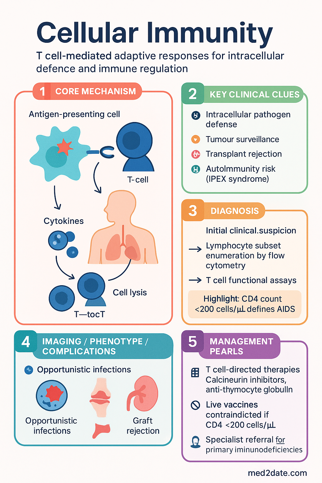
- Cellular immunity — T cell-mediated adaptive responses — is essential for defence against intracellular pathogens (mycobacteria, fungi, viruses), tumour surveillance, and transplant rejection.
- CD4⁺ helper T cells coordinate immunity through cytokine release: Th1 (cellular), Th2 (humoral/allergic), Th17 (mucosal), Tfh (germinal centre), and Treg (suppression).
- CD8⁺ cytotoxic T lymphocytes (CTLs) kill via perforin/granzyme exocytosis and Fas/FasL signalling — critical for viral clearance and anti-tumour immunity.
- Memory T cells (Tcm, Tem, Trm) provide rapid recall responses; Trm cells reside in mucosal tissue and skin for frontline defence.
- Regulatory T cells (Tregs, CD4⁺CD25⁺FOXP3⁺) maintain self-tolerance; deficiency causes IPEX syndrome and multi-organ autoimmunity.
- Lymphocyte subset enumeration (CD3, CD4, CD8, CD19, CD16/56) by flow cytometry is MBS-listed (Item 69332) and essential for immunodeficiency workup.
- Absolute CD4 count remains the principal marker for HIV staging and opportunistic infection risk; <200 cells/µL defines AIDS.
- T cell functional assays (PHA/anti-CD3 lymphocyte proliferation, intracellular cytokine staining) are available at tertiary centres and reference labs.
- Primary immunodeficiencies affecting T cells (DiGeorge, SCID, STAT3/STAT1 GOF, IPEX) require urgent immunology referral for haematopoietic stem cell transplant assessment.
- Secondary T cell dysfunction is most commonly caused by HIV, immunosuppressive therapy (corticosteroids, calcineurin inhibitors, biologics), malnutrition, and haematological malignancy.
- T cell-directed therapies include calcineurin inhibitors (cyclosporin, tacrolimus), anti-thymocyte globulin, anti-CD52 (alemtuzumab), and emerging checkpoint inhibitors (anti-PD-1/PD-L1, anti-CTLA-4).
- Aboriginal and Torres Strait Islander peoples have higher rates of infections requiring robust cellular immunity (TB, invasive pneumococcal disease, rheumatic fever); culturally safe immunology referral pathways are essential.
- Live vaccines (MMR, varicella, BCG, yellow fever) are contraindicated when CD4 <200 cells/µL or in severe T cell immunodeficiency — assess prior to vaccination.
- Flow cytometry turnaround in most Australian public hospital labs is 3–5 business days; specialist lymphocyte function testing may take 2–4 weeks.
Introduction & Australian Epidemiology
Cellular immunity — the arm of adaptive immunity mediated by T lymphocytes — is central to host defence against intracellular pathogens, tumour surveillance, graft rejection, and regulation of the broader immune response. Unlike humoral (antibody-mediated) immunity, cellular immunity requires direct cell-to-cell contact and cytokine-mediated signalling to eliminate infected or malignant cells and to coordinate downstream effector functions.
T cells originate from haematopoietic precursors in the bone marrow, undergo positive and negative selection in the thymus, and emerge as naïve CD4⁺ or CD8⁺ single-positive cells bearing unique T cell receptors (TCRs). Upon antigen encounter in secondary lymphoid organs, naïve T cells differentiate into effector and memory subsets that mediate both immediate and long-term protection.
The Australian landscape for cellular immunology is shaped by several factors:
- HIV programme: Australia's universal access to antiretroviral therapy (ART) through the PBS Pharmaceutical Benefits Scheme has achieved viral suppression in >95% of those on treatment, preserving CD4 counts and reducing opportunistic infections.
- Transplant immunology: National transplant programmes rely on calcineurin inhibitor-based regimens; tertiary centres perform HLA typing and donor-specific antibody (DSA) monitoring with virtual crossmatching.
- Primary immunodeficiency: The Australian National Immunodeficiency Registry estimates a prevalence of 1 in 1,200 for combined variable immunodeficiency (CVID) and related disorders; severe T cell deficiencies (SCID, DiGeorge) are identified through newborn screening programmes now operational in NSW, ACT, and expanding nationally.
- Checkpoint immunotherapy: Immune checkpoint inhibitors (nivolumab, pembrolizumab, ipilimumab) are PBS-listed for melanoma, NSCLC, renal cell carcinoma, and other malignancies; immune-related adverse events (irAEs) from T cell over-activation are managed by multidisciplinary teams.
- ATSI health: Aboriginal and Torres Strait Islander peoples experience higher rates of conditions requiring intact cellular immunity, including TB, rheumatic heart disease, and invasive Group A Streptococcus disease.
Core Concepts in T Cell Biology
The T cell compartment is defined by surface markers and functional capacity:
| T Cell Subset | Surface Markers | Primary Function | Key Cytokines / Molecules |
|---|---|---|---|
| CD4⁺ Naïve | CD3⁺CD4⁺CD45RA⁺CCR7⁺ | Precursor to helper subsets | IL-7 (survival), TCR engagement required for activation |
| Th1 | CD3⁺CD4⁺IFN-γ⁺ | Intracellular pathogen defence, macrophage activation | IFN-γ, TNF-α, IL-2; T-bet transcription factor |
| Th2 | CD3⁺CD4⁺IL-4⁺ | Helminth defence, B cell class switching to IgE | IL-4, IL-5, IL-13; GATA3 transcription factor |
| Th17 | CD3⁺CD4⁺IL-17A⁺ | Mucosal defence, neutrophil recruitment | IL-17A, IL-17F, IL-22; RORγt transcription factor |
| Tfh | CD3⁺CD4⁺CXCR5⁺PD-1⁺ | Germinal centre B cell help, antibody affinity maturation | IL-21, ICOS, CD40L; Bcl-6 transcription factor |
| Treg | CD3⁺CD4⁺CD25⁺FOXP3⁺ | Immune suppression, tolerance | IL-10, TGF-β, CTLA-4, IL-35 |
| CD8⁺ CTL | CD3⁺CD8⁺ | Killing of virus-infected and tumour cells | Perforin, granzymes, IFN-γ, FasL |
| MAIT | CD3⁺CD161⁺Vα7.2⁺ | Mucosal innate-like defence against bacteria | IFN-γ, IL-17; MR1-restricted |
T Cell Subsets
Thymic Development & Selection
T cell precursors migrate from the bone marrow to the thymus, where they undergo a tightly regulated maturation programme:
Effector T Cell Subsets in Peripheral Blood
Once in the periphery, naïve T cells encounter antigen on antigen-presenting cells (APCs) in secondary lymphoid organs and differentiate into effector subsets. The prevailing cytokine milieu during activation determines subset commitment:
| Subset | Activating Cytokines | Key Transcription Factor | Effector Cytokines | Target Pathogens / Roles | Dysregulation |
|---|---|---|---|---|---|
| Th1 | IL-12, IFN-γ | T-bet | IFN-γ, TNF-α, IL-2 | Mycobacteria, viruses, intracellular bacteria | Autoimmunity (MS, T1DM, RA) |
| Th2 | IL-4 | GATA3 | IL-4, IL-5, IL-13 | Helminths | Asthma, atopic dermatitis, allergic rhinitis |
| Th17 | IL-6 + TGF-β (mice); IL-1β, IL-6, IL-23 (humans) | RORγt | IL-17A, IL-17F, IL-22 | Extracellular bacteria (Klebsiella, Staph), fungi (Candida) | Psoriasis, IBD, ankylosing spondylitis |
| Tfh | IL-6, IL-21, ICOS | Bcl-6 | IL-21, IL-4 | Germinal centre reactions, high-affinity antibody | SLE, angioimmunoblastic T cell lymphoma |
| Treg | TGF-β, IL-2 | FOXP3 | IL-10, TGF-β, IL-35 | Immune suppression, self-tolerance | IPEX syndrome, autoimmunity |
Unconventional T Cells
Beyond classical αβ T cells, several unconventional T cell populations contribute to immune defence:
- γδ T cells: Constitute 1–5% of peripheral blood T cells; enriched in mucosal tissues. Recognise phosphoantigens and stress-induced ligands (MICA/MICB) without classical MHC restriction. Important in defence against mycobacteria and epithelial tumours.
- MAIT cells: Mucosal-associated invariant T cells recognise microbial vitamin B metabolites presented by MR1. Abundant in human blood (1–10% of T cells) and liver. Rapidly produce IFN-γ and IL-17 upon bacterial encounter. Depleted in HIV infection despite ART.
- NKT cells: Recognise glycolipid antigens presented by CD1d. Invariant NKT (iNKT) cells produce large cytokine bursts early in infection. Deficiency associated with increased susceptibility to certain infections.
- Double-negative (DN) T cells: CD3⁺CD4⁻CD8⁻ T cells that can suppress autoreactive T cells and are expanded in autoimmune lymphoproliferative syndrome (ALPS).
CD4⁺ Helper T Cell Functions
CD4⁺ T helper cells are the conductors of adaptive immunity. Their functions extend far beyond simple cytokine production — they direct the type, magnitude, and location of immune responses. Understanding CD4⁺ T cell biology is essential for managing HIV infection, autoimmune diseases, transplant rejection, and vaccine design.
Mechanisms of Help
Tfh cells in germinal centres provide signals to B cells via CD40L–CD40 interaction and IL-21 secretion, driving class-switch recombination, somatic hypermutation, and affinity maturation. Without Tfh help, germinal centre reactions fail, and long-lived plasma cells and memory B cells do not form.
Clinical correlate: Patients with Hyper-IgM syndrome (CD40L deficiency) have absent class switching and are susceptible to Pneumocystis jirovecii and Cryptosporidium.
IFN-γ produced by Th1 cells activates macrophages, enhancing phagolysosome fusion, reactive oxygen/nitrogen species production, and MHC-II expression. This classical activation is critical for killing intracellular pathogens such as Mycobacterium tuberculosis and Leishmania.
Clinical correlate: Patients on anti-TNF therapy (infliximab, adalimumab) have impaired granuloma maintenance and risk of TB reactivation.
CD4⁺ T Cell Roles in Specific Diseases
CD4 Count Interpretation in Australian Practice
| CD4 Count (cells/µL) | Risk Level | Clinical Implications | Prophylaxis Required |
|---|---|---|---|
| 500–1,500 (normal) | Normal | Adequate immune function | Standard vaccination schedule |
| 350–500 | Mild reduction | ART initiation recommended (all PLHIV); increased infection risk minimal | Nil additional |
| 200–350 | Moderate reduction | Increased risk of TB, oral candidiasis, herpes zoster, bacterial pneumonia | TB screening (IGRA/TST); consider Bactrim if CD4 approaching 200 |
| 100–200 | Severe (AIDS if <200) | PCP, oesophageal candidiasis, disseminated HSV | Co-trimoxazole 480 mg daily (PCP prophylaxis) |
| 50–100 | Very severe | Toxoplasma encephalitis, Cryptococcal meningitis, disseminated MAC | Co-trimoxazole + azithromycin 1,200 mg weekly (MAC prophylaxis if CD4 <50) |
| <50 | Critical | CMV retinitis, disseminated MAC, CNS lymphoma (EBV-driven) | Full opportunistic infection prophylaxis; ophthalmology review |
Therapeutic Modulation of CD4⁺ T Cells
Multiple drug classes target CD4⁺ T cell function in transplantation, autoimmunity, and lymphoma:
CD8⁺ Cytotoxic Mechanisms
CD8⁺ cytotoxic T lymphocytes (CTLs) are the primary effectors for eliminating virus-infected cells, intracellular bacteria-harbouring cells, and tumour cells. CTL killing is exquisitely specific — each CTL recognises its cognate peptide presented on MHC class I molecules, which are expressed on virtually all nucleated cells.
Killing Mechanisms
CTLs employ two major cytotoxic pathways:
- Upon TCR engagement, the immunological synapse forms and lytic granules polarise toward the target cell.
- Perforin polymerises in the target cell membrane, forming pores (~16 nm diameter).
- Granzyme B enters through perforin pores and directly cleaves caspase-3, -7, and -10, triggering apoptosis.
- Granzyme B also cleaves Bid → tBid → mitochondrial outer membrane permeabilisation → cytochrome c release → caspase-9 activation (intrinsic apoptotic pathway).
- Families with biallelic PRF1 mutations (perforin deficiency) develop familial haemophagocytic lymphohistiocytosis (FHL type 2) — a life-threatening hyperinflammatory syndrome.
- Fas ligand (CD95L, TNFSF6) on CTLs engages Fas (CD95, TNFRSF6) on target cells.
- Fas trimerisation recruits FADD and procaspase-8 to form the death-inducing signalling complex (DISC).
- Caspase-8 activation → cleavage of caspase-3 → apoptosis.
- Important for immune contraction (activation-induced cell death, AICD) and liver immunopathology.
- Mutations in FAS or FASL cause autoimmune lymphoproliferative syndrome (ALPS) — characterised by lymphadenopathy, splenomegaly, and accumulation of double-negative T cells.
Non-Cytolytic Functions of CD8⁺ T Cells
Beyond direct killing, CTLs contribute to immunity through:
- IFN-γ production: Enhances MHC-I expression on neighbouring cells, upregulates antigen processing machinery, and activates macrophages.
- TNF-α secretion: Promotes inflammation, endothelial activation, and has direct anti-viral and anti-tumour effects.
- Cytotoxic degranulation without killing: Partial degranulation can deliver granzymes that induce non-apoptotic effects (pyroptosis via gasdermin E cleavage in some contexts).
- Immunosurveillance: Tissue-resident memory CD8⁺ T cells (Trm) positioned in epithelia provide rapid responses to re-infection at barrier sites.
CD8⁺ T Cells in Key Clinical Scenarios
| Clinical Scenario | CD8⁺ Role | Australian Relevance |
|---|---|---|
| HIV infection | HIV-specific CTLs suppress viraemia (elite controllers); escape mutations in immunodominant epitopes drive viral evolution | ~29,000 PLHIV; CD8 responses correlate with set-point viral load |
| SARS-CoV-2 | Robust CD8⁺ responses correlate with mild disease; impaired responses in severe COVID-19 | T cell immunity persists after antibody waning; relevant to booster strategy |
| Solid organ transplant | Donor-specific CD8⁺ T cells mediate acute cellular rejection (ACR); controlled by calcineurin inhibitors and anti-metabolites | ~1,500 transplants/year; protocol biopsies monitor for ACR |
| Melanoma | Tumour-infiltrating lymphocytes (TILs) with CD8⁺ predominance predict response to checkpoint inhibitors | Australia has highest melanoma incidence globally; checkpoint immunotherapy PBS-listed |
| CMV reactivation | CMV-specific CD8⁺ T cells control latent CMV; depletion in transplant/HIV leads to reactivation | CMV PCR monitoring post-transplant standard in Australian centres |
| Hepatitis B | HBV-specific CTL responses determine clearance vs. chronicity; dysfunctional/exhausted in chronic HBV | ~220,000 people living with chronic HBV in Australia; higher prevalence in ATSI and CALD communities |
Checkpoint Inhibitors & CD8⁺ T Cell Reinvigoration
Immune checkpoint inhibitors (ICIs) reinvigorate exhausted CD8⁺ T cells in the tumour microenvironment:
Immune-related adverse events (irAEs) from checkpoint inhibitors reflect unrestrained T cell activation against self-antigens. Common irAEs include colitis, hepatitis, pneumonitis, thyroiditis, hypophysitis, and dermatitis. Severe irAEs (Grade 3–4) require high-dose corticosteroids (methylprednisolone 1–2 mg/kg IV) and may necessitate infliximab (colitis) or mycophenolate (hepatitis).
Memory & Regulation
Memory T Cell Subsets
Immunological memory is the foundation of vaccination and long-term pathogen protection. Memory T cells persist for decades and provide faster, stronger, and qualitatively superior responses upon antigen re-encounter.
| Memory Subset | Surface Markers | Location | Function | Lifespan |
|---|---|---|---|---|
| Tcm (Central memory) | CD45RO⁺CCR7⁺CD62L⁺ | Secondary lymphoid organs (lymph nodes, spleen) | High proliferative capacity upon re-stimulation; self-renewing via IL-7/IL-15 | Decades |
| Tem (Effector memory) | CD45RO⁺CCR7⁻CD62L⁻ | Peripheral tissues, blood | Rapid effector function (cytokine production, cytotoxicity) upon re-encounter; less proliferative than Tcm | Years to decades |
| Trm (Tissue-resident memory) | CD69⁺CD103⁺ (variable) | Non-lymphoid tissues (skin, lung, gut, reproductive tract) | First-line barrier defence; do not recirculate; produce IFN-γ and TNF-α rapidly; recruit circulating immune cells | Years (self-maintained locally) |
| Tscm (Stem cell memory) | CD45RA⁺CCR7⁺CD62L⁺CD95⁺CD122⁺ | Blood, lymphoid organs | Stem-like self-renewal; can differentiate into Tcm, Tem, and effector cells; rare population (~2–3% of CD4⁺) | Lifetime |
Maintenance of Memory T Cells
Memory T cell persistence depends on:
- Homeostatic cytokines: IL-7 (produced by stromal cells) and IL-15 (produced by DCs and macrophages) drive slow homeostatic proliferation of memory T cells, maintaining the pool without antigen stimulation.
- Metabolic reprogramming: Memory T cells shift from glycolysis (effector cells) to fatty acid oxidation and oxidative phosphorylation, enabling long-term survival with minimal nutrient requirements.
- Epigenetic imprinting: Memory T cells carry permissive chromatin marks at effector gene loci, allowing rapid transcription upon restimulation (poised chromatin state).
- Antigen-independent signals: MHC-II interactions (for CD4⁺ memory) and tonic TCR signalling contribute to memory maintenance.
Regulatory T Cells (Tregs)
Tregs are indispensable for maintaining immune homeostasis and preventing autoimmunity. They constitute 5–10% of circulating CD4⁺ T cells in healthy adults.
Develop in the thymus during negative selection. High-affinity TCR engagement with self-antigen/MHC on mTECs drives FOXP3 expression and commitment to the Treg lineage. These cells are critical for dominant tolerance — actively suppressing autoreactive T cells that escape deletion.
Marker profile: CD4⁺CD25^(hi)CD127^(lo)FOXP3⁺Helios⁺Neuropilin-1⁺
Induced in the periphery from naïve CD4⁺ T cells by TGF-β and retinoic acid, particularly in mucosal tissues (gut). Essential for tolerance to commensal flora, food antigens, and fetal antigens during pregnancy.
Marker profile: CD4⁺CD25^(hi)CD127^(lo)FOXP3⁺Helios⁻Neuropilin-1⁻
Mechanisms of Treg Suppression
- IL-2 consumption: Tregs express high-affinity IL-2Rα (CD25) but do not produce IL-2, acting as an "IL-2 sink" that deprives effector T cells of this essential growth factor.
- CTLA-4-mediated suppression: Constitutive CTLA-4 expression on Tregs strips CD80/CD86 from APCs (trans-endocytosis), reducing co-stimulation for effector T cells.
- Immunosuppressive cytokines: Secretion of IL-10, TGF-β, and IL-35 inhibits APC function and effector T cell proliferation.
- Granzyme/perforin: Some Tregs can directly kill effector T cells and APCs via cytolytic mechanisms.
- Metabolic disruption: CD39/CD73 ectoenzymes on Tregs convert ATP to adenosine, which suppresses effector T cell function via A2A receptor signalling.
- Dendritic cell modulation: Tregs induce tolerogenic DCs via LAG-3–MHC-II interaction, promoting anergy in subsequently activated T cells.
Treg Dysfunction in Disease
Clinical Assessment of Memory & Regulatory T Cells
In Australian clinical practice, assessment of T cell memory and regulatory compartments is performed by:
- Flow cytometry (MBS Item 69332): Lymphocyte subset panel includes CD45RA/RO and CCR7 to delineate naïve, Tcm, and Tem populations. CD25/CD127 ratio approximates Treg frequency (FOXP3 intracellular staining confirmatory).
- TREC (T cell receptor excision circle) assay: Measures thymic output; low TRECs indicate reduced naïve T cell production (DiGeorge, post-chemotherapy, ageing). Available at select reference labs.
- TCR repertoire analysis: Next-generation sequencing of TCRβ CDR3 regions assesses clonality and diversity. Expanded clones may indicate chronic infection, malignancy, or alloreactivity. Available through specialised genomics services.
- Vaccine response testing: T cell responses to recall antigens (tetanus, CMV, EBV) by ELISpot (IFN-γ) confirm functional immunological memory. Available at Westmead, RMH, and SA Pathology.
Pathophysiology
Disorders of cellular immunity arise from defects at multiple levels of T cell biology — from thymic development through effector function and regulation. Understanding the pathophysiology is essential for targeted diagnosis and management.
Classification of T Cell Immunodeficiencies
| Category | Examples | Pathophysiological Basis | Inheritance |
|---|---|---|---|
| Combined immunodeficiencies | Severe combined immunodeficiency (SCID): X-linked (IL2RG), ADA deficiency, Artemis, RAG1/2 | Absent or profoundly reduced T cells ± B/NK cells; failed V(D)J recombination or cytokine signalling | X-linked, AR |
| Thymic defects | DiGeorge syndrome (22q11.2 deletion) | Thymic hypoplasia/aplasia → reduced T cell production; severity proportional to thymic tissue present | Sporadic (de novo) or AD |
| Predominantly T cell defects | CD3 deficiency, ZAP-70 deficiency, MHC-II deficiency (Bare lymphocyte syndrome) | Defective TCR signalling or antigen presentation | AR |
| Immune dysregulation | IPEX (FOXP3), ALPS (FAS/FASL/CASP10), CTLA-4 haploinsufficiency | Loss of peripheral tolerance; uncontrolled T cell proliferation or impaired apoptosis | X-linked (IPEX), AD/AR (ALPS) |
| Syndromic immunodeficiencies | Ataxia-telangiectasia (ATM), Wiskott-Aldrich (WASP), CHARGE syndrome | DNA repair defects, cytoskeletal dysfunction, or developmental genes affecting multiple lineages | AR (AT, WAS), AD (CHARGE) |
| Secondary (acquired) | HIV/AIDS, iatrogenic (corticosteroids, calcineurin inhibitors, anti-CD20, biologics), malnutrition, malignancy | Direct T cell destruction, impaired production, or functional suppression | N/A |
T Cell Exhaustion
Chronic antigen stimulation (persistent viral infection, tumour) drives T cell exhaustion — a state of progressive loss of effector function characterised by:
- Upregulation of inhibitory receptors: PD-1, LAG-3, TIM-3, TIGIT, CTLA-4
- Hierarchical loss of cytokine production: IL-2 first, then TNF-α, then IFN-γ (functional avidity decreases)
- Reduced proliferative capacity and cytotoxicity
- Distinct epigenetic landscape (TOX transcription factor drives exhaustion programme)
- Presence of progenitor exhausted cells (TCF1⁺PD-1⁺) that can be reinvigorated — the target of checkpoint immunotherapy
Investigations
Assessment of cellular immunity involves quantification of T cell populations, assessment of T cell function, and evaluation of underlying aetiology. Investigation selection depends on clinical context.