📋 Key Information Summary
- Antigen presenting cells (APCs) process and display peptide fragments on MHC molecules to activate T lymphocytes — the central event bridging innate and adaptive immunity.
- Professional APCs — dendritic cells (DCs), macrophages, and B cells — constitutively express MHC class II and are the primary activators of naïve CD4⁺ T cells.
- Dendritic cells are the most potent professional APCs and uniquely capable of priming naïve T cells, making them critical for initiating primary immune responses and vaccine efficacy.
- Macrophages serve dual roles as APCs and effector phagocytes; they are prominent in granulomatous inflammation and intracellular pathogen defence (e.g., Mycobacterium tuberculosis).
- B cells use the B-cell receptor (BCR) to capture specific antigen at very low concentrations, making them efficient APCs during secondary immune responses and in autoimmune disease.
- APC maturation involves upregulation of MHC II, co-stimulatory molecules (CD80/CD86), and cytokine production; immature APCs promote tolerance, while mature APCs drive activation.
- T-cell activation requires two signals: Signal 1 (TCR–MHC/peptide) and Signal 2 (CD28–B7 co-stimulation). Without Signal 2, T cells become anergic — the basis of peripheral tolerance.
- A third signal (polarising cytokines such as IL-12, IL-4, IL-6, TGF-β) from APCs determines the Th1/Th2/Th17/Treg differentiation fate of activated T cells.
- APC dysfunction underlies numerous clinical conditions: immunodeficiency (e.g., bare lymphocyte syndrome type II), autoimmunity, transplant rejection, and tumour immune evasion.
- Regulatory mechanisms including CTLA-4, PD-1/PD-L1, and indoleamine 2,3-dioxygenase (IDO) in APCs maintain peripheral tolerance; dysregulation contributes to autoimmune disease in Australians.
- Understanding APC biology informs clinical decisions in vaccination strategy, immunosuppression (calcineurin inhibitors block APC-dependent T-cell activation), and emerging immunotherapies (DC vaccines, checkpoint inhibitors).
- Aboriginal and Torres Strait Islander Australians experience higher burdens of infections and autoimmune conditions where APC function is critical; culturally safe immunological education is essential.
- Current Australian research at WEHI, the Doherty Institute, and Garvan Institute continues to elucidate APC biology for translation into vaccines, cancer immunotherapy, and tolerance induction.
Introduction & Australian Immunological Context
Antigen presenting cells (APCs) are specialised immune cells that capture, process, and display foreign and self-peptides on major histocompatibility complex (MHC) molecules to T lymphocytes. In doing so, they function as the critical bridge between innate and adaptive immunity, initiating, shaping, and regulating the specificity and magnitude of the T-cell response. Without effective antigen presentation, adaptive immunity cannot be appropriately primed, leaving the host susceptible to infection, malignancy, or — paradoxically — autoimmune pathology when presentation becomes dysregulated.
The concept of antigen presentation was first elucidated in the 1970s and 1980s through the work of Zinkernagel and Doherty (Nobel Prize, 1996), who demonstrated MHC-restricted antigen recognition at the John Curtin School of Medical Research in Canberra — a landmark contribution to global immunology from an Australian institution. Subsequent decades of research have defined multiple APC subsets, their maturation pathways, co-stimulatory requirements, and tolerance-promoting functions.
In clinical practice across Australia, APC biology underpins vaccination programmes, management of immunodeficiency states, transplant immunology, cancer immunotherapy, and the understanding of autoimmune diseases that disproportionately affect Australians — particularly type 1 diabetes, multiple sclerosis, rheumatoid arthritis, and inflammatory bowel disease. A thorough understanding of APC function is therefore essential for clinicians managing these conditions in primary and specialist care.
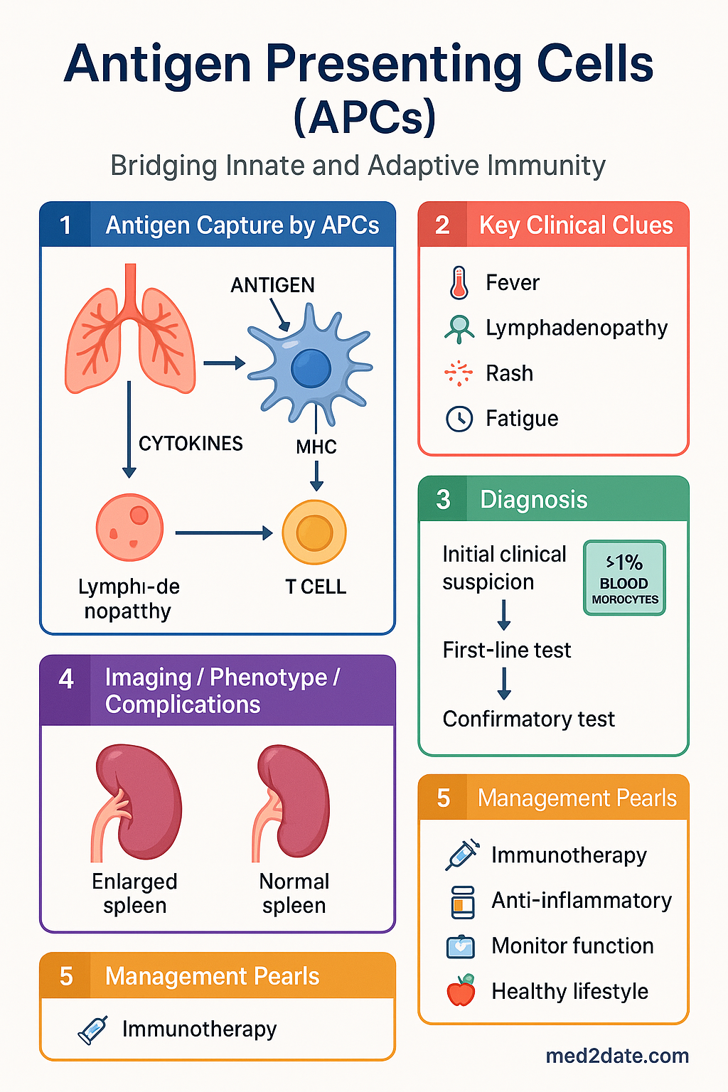
Types of Antigen Presenting Cells
All nucleated cells express MHC class I molecules and can present endogenous (intracellular) peptides to CD8⁺ cytotoxic T cells — a process termed cross-presentation when performed by specialised APCs on exogenous antigens. However, the term "professional APC" is reserved for cells that constitutively express MHC class II and co-stimulatory molecules, enabling activation of naïve CD4⁺ helper T cells. The three principal professional APCs are dendritic cells, macrophages, and B lymphocytes.
| Feature | Dendritic Cells | Macrophages | B Lymphocytes |
|---|---|---|---|
| MHC II expression | Constitutive (high, upregulated with maturation) | Constitutive (inducible by IFN-γ) | Constitutive (moderate) |
| Co-stimulation (CD80/86) | High (after maturation) | Moderate (induced by TLR ligands) | Moderate (after activation) |
| Antigen capture mechanism | Macropinocytosis, receptor-mediated endocytosis, phagocytosis | Phagocytosis, FcγR, complement receptors | BCR-mediated (antigen-specific) |
| Primary role | Initiation of naïve T-cell responses | Effector function + antigen presentation during inflammation | Focused antigen presentation in secondary responses |
| Cross-presentation | Yes — key function (cDC1 subset) | Limited | Rare |
| Location (lymph node) | T-cell zones (paracortex) | Subcapsular sinus, medullary cords | B-cell follicles, germinal centres |
Dendritic Cells
Dendritic cells (DCs) are the most potent professional APCs and are uniquely capable of activating naïve T cells — a property that places them at the apex of the immune response hierarchy. They originate from bone marrow precursors and exist in two broad functional states: immature DCs residing in peripheral tissues (e.g., Langerhans cells in the epidermis) and mature DCs in secondary lymphoid organs.
DC subsets include:
- Conventional DC type 1 (cDC1): Specialised in cross-presentation of exogenous antigens on MHC class I, driving CD8⁺ cytotoxic T-cell responses critical for anti-tumour and anti-viral immunity. Depend on transcription factors BATF3 and IRF8. Key for checkpoint inhibitor efficacy in cancer.
- Conventional DC type 2 (cDC2): Present antigens on MHC class II, primarily activating CD4⁺ T helper cells. Drive Th1, Th2, and Th17 responses depending on the cytokine milieu. Depend on IRF4.
- Plasmacytoid DCs (pDCs): Produce large quantities of type I interferons (IFN-α/β) in response to viral nucleic acids via TLR7 and TLR9. Less efficient as APCs but critical in anti-viral defence.
- Monocyte-derived DCs (moDCs): Differentiate from monocytes during inflammation; abundant at sites of infection and in the synovium of patients with rheumatoid arthritis.
Macrophages
Macrophages are tissue-resident phagocytes derived from embryonic precursors or circulating monocytes. While their primary function is innate effector activity — phagocytosis, microbicidal killing, and cytokine secretion — they also serve as APCs, particularly in the context of ongoing inflammation and in granulomatous disease.
Macrophage polarisation states relevant to APC function:
- M1 (classically activated): Induced by IFN-γ and TLR ligands; produce IL-12, TNF-α, and reactive oxygen/nitrogen species. Promote Th1 responses. Critical in intracellular pathogen defence (Mycobacterium tuberculosis, Leishmania).
- M2 (alternatively activated): Induced by IL-4 and IL-13; produce IL-10 and TGF-β. Promote Th2 responses, tissue repair, and fibrosis. Relevant in helminth infections and allergic disease.
In tuberculosis — which has a significant burden in Aboriginal and Torres Strait Islander communities in northern Australia — macrophage APC function is central to granuloma formation and disease containment. Impaired macrophage presentation contributes to disease progression in immunocompromised patients.
B Lymphocytes
B cells function as specialised APCs that use their clonally rearranged B-cell receptor (BCR) to capture specific antigen with extremely high affinity. This allows B cells to concentrate and present antigen at concentrations 1,000–10,000-fold lower than required by DCs or macrophages.
B-cell APC function is particularly important in:
- Secondary immune responses: Memory B cells efficiently present recall antigens to memory T cells in germinal centres.
- Autoimmune disease: In rheumatoid arthritis and systemic lupus erythematosus, autoreactive B cells present self-antigens to T cells, perpetuating the autoimmune cycle. Rituximab (anti-CD20) depletes B cells and is PBS-listed for refractory RA in Australia.
- Viral infections: B cells capture viral particles via BCR and present them to T follicular helper cells, driving affinity maturation and class switching.
Maturation & Activation
APC maturation is the critical transition from a tolerogenic resting state to an immunogenic activated state. This process is triggered by pathogen-associated molecular patterns (PAMPs) and damage-associated molecular patterns (DAMPs) acting on pattern recognition receptors (PRRs), including Toll-like receptors (TLRs), NOD-like receptors (NLRs), RIG-I-like receptors (RLRs), and C-type lectin receptors.
Cross-Presentation
Cross-presentation is the ability of certain APCs (primarily cDC1 dendritic cells) to load exogenous antigens onto MHC class I molecules, thereby activating CD8⁺ T cells against pathogens or tumours that do not directly infect APCs. This process is essential for anti-tumour immunity and is a major target of cancer immunotherapy strategies. The cytosolic pathway involves antigen escape from endosomes into the cytosol, proteasomal degradation, and TAP-dependent loading onto MHC I in the ER — mirroring the classical MHC I pathway for endogenous antigens.
Co-stimulatory Signals
T-cell activation requires the integration of multiple signals delivered by APCs. The two-signal model, first proposed by Bretscher and Cohn (1970) and later refined by Lafferty and Cunningham (1975), remains the foundational framework for understanding APC–T-cell interaction.
Key Co-stimulatory and Co-inhibitory Receptor Pairs
| APC Ligand | T-cell Receptor | Effect | Clinical Relevance |
|---|---|---|---|
| CD80 / CD86 (B7-1/B7-2) | CD28 | Co-stimulation (activation) | Basis of T-cell activation; blocked by abatacept (CTLA-4-Ig, Orencia®) in RA |
| CD80 / CD86 | CTLA-4 | Co-inhibition (brake) | Higher affinity than CD28; ipilimumab (anti-CTLA-4) used in melanoma (PBS-authority) |
| PD-L1 / PD-L2 | PD-1 | Co-inhibition | Nivolumab, pembrolizumab (anti-PD-1) — PBS-listed for melanoma, NSCLC, RCC |
| CD40 | CD40L (CD154) | Bidirectional — enhances APC function and T-cell help | Critical for germinal centre reactions, B-cell class switching, DC licensing for cross-presentation |
| ICOS-L | ICOS | T follicular helper cell development | Germinal centre function; deficiency causes common variable immunodeficiency (CVID) |
| OX40L | OX40 (CD134) | T-cell survival and memory | Target of experimental cancer immunotherapies in Australian trials |
Negative Co-stimulation & Immune Checkpoints
Co-inhibitory receptors act as immune checkpoints that prevent excessive T-cell activation and maintain self-tolerance. CTLA-4 is upregulated after T-cell activation and competes with CD28 for B7 ligands with ~20-fold higher affinity. PD-1 is induced on activated T cells and engages PD-L1/PD-L2 expressed by APCs, tumour cells, and stromal cells to deliver an inhibitory signal. Both pathways are exploited therapeutically in Australian cancer care: ipilimumab (anti-CTLA-4) and nivolumab/pembrolizumab (anti-PD-1) are PBS-listed for multiple malignancies under authority prescription.
Lag-3, TIM-3, TIGIT, and VISTA represent emerging co-inhibitory receptors under investigation in combination strategies at Australian cancer centres.
Role in Tolerance
APCs play a dual role: they are essential for immunity but equally critical for establishing and maintaining immune tolerance. Tolerance operates through two main mechanisms — central tolerance (in the thymus and bone marrow) and peripheral tolerance (in tissues and lymph nodes) — and APCs are integral to both.
Central Tolerance
In the thymus, medullary thymic epithelial cells (mTECs) express the transcription factor AIRE (autoimmune regulator), which drives ectopic expression of thousands of tissue-restricted antigens. Developing thymocytes that strongly recognise self-peptide–MHC complexes on mTECs and thymic DCs undergo negative selection (clonal deletion). Mutations in AIRE cause autoimmune polyendocrinopathy-candidiasis-ectodermal dystrophy (APECED/APS-1), a rare condition seen in Australian paediatric immunology clinics.
Peripheral Tolerance
Peripheral tolerance mechanisms in which APCs participate include:
Tolerogenic Dendritic Cells — Therapeutic Potential
Tolerogenic DCs (tolDCs) are generated in vitro by exposing monocyte-derived DCs to immunosuppressive agents (dexamethasone, vitamin D₃, rapamycin) or IL-10. These tolDCs express low MHC II, low CD80/86, and high IL-10/TGF-β, and can induce antigen-specific Tregs and anergy. Clinical trials in rheumatoid arthritis, type 1 diabetes, and solid organ transplantation are underway globally, with Australian centres participating in early-phase studies.
Clinical Relevance for Australian Practice
APC Dysfunction in Immunodeficiency
Primary immunodeficiencies affecting APC function are rare but clinically significant:
- Bare lymphocyte syndrome type II (BLS II): Mutations in MHC class II transactivator (CIITA) or promoter elements result in absent MHC class II expression. Patients present in infancy with severe recurrent infections, chronic diarrhoea, and failure to thrive. Diagnosis involves flow cytometry for MHC II expression (available at major Australian immunology centres). Haematopoietic stem cell transplant is the only curative therapy.
- IRF8 deficiency: Loss-of-function mutations in IRF8 abolish DC and monocyte development, causing severe immunodeficiency in infancy.
- GATA2 deficiency: Monocytopenia and DC deficiency predispose to atypical mycobacterial infections, viral infections (HPV), and myelodysplasia. Seen in Australian adult immunology clinics.
- STAT3 loss-of-function (Hyper-IgE syndrome): Impaired Th17 differentiation due to defective APC-derived IL-6/IL-23 signalling, predisposing to mucocutaneous candidiasis and staphylococcal infections.
APCs in Transplant Immunology
APCs are central to allograft rejection through direct and indirect allorecognition:
- Direct recognition: Recipient T cells recognise intact donor MHC molecules on donor APCs (passenger leucocytes) within the graft. This drives acute rejection and is the primary target of calcineurin inhibitors (tacrolimus, ciclosporin — PBS-listed).
- Indirect recognition: Recipient APCs process shed donor MHC molecules and present donor-derived peptides on self-MHC to recipient T cells. This pathway predominates in chronic rejection.
- Semi-direct recognition: Recipient APCs acquire intact donor MHC molecules via extracellular vesicle transfer (trogocytosis), combining features of both pathways.
Immunosuppressive agents used in Australian transplant programmes target APC-dependent pathways: tacrolimus and ciclosporin calcineurin inhibitors block NFAT-dependent IL-2 transcription; mycophenolate inhibits purine synthesis in T and B cells; and belatacept (CTLA-4-Ig) blocks CD80/86 co-stimulation directly.
APCs in Vaccine Immunology
Vaccine efficacy depends on effective APC activation and antigen presentation. Adjuvants used in the Australian National Immunisation Programme enhance DC maturation:
- Aluminium salts: Activate NLRP3 inflammasome, promote DC maturation and Th2 skewing (used in Infanrix®, Prevenar 13®, Boostrix®).
- AS01 (MPL + QS-21): Activates DCs via TLR4 (MPL) and promotes cross-presentation (used in Shingrix®, Mosquirix®).
- mRNA vaccines: Lipid nanoparticles are taken up by DCs at the injection site; mRNA translation produces antigen intracellularly for MHC I and MHC II presentation (Comirnaty®, Spikevax® — COVID-19 programme).
Special Populations
Aboriginal and Torres Strait Islander Health Considerations
📚 References
- 1. Zinkernagel RM, Doherty PC. Restriction of in vitro T cell-mediated cytotoxicity in lymphocytic choriomeningitis within a syngeneic or semiallogeneic system. Nature. 1974;248(5450):701–702.
- 2. Banchereau J, Steinman RM. Dendritic cells and the control of immunity. Nature. 1998;392(6673):245–252.
- 3. Sharpe AH, Freeman GJ. The B7-CD28 superfamily. Nature Reviews Immunology. 2002;2(2):116–126.
- 4. Merad M, Sathe P, Helft J, Miller J, Mortha A. The dendritic cell lineage: ontogeny and function of dendritic cells and their subsets in the steady state and the inflamed setting. Annual Review of Immunology. 2013;31:563–604.
- 5. Murray PJ, Allen JE, Biswas SK, et al. Macrophage activation and polarization: nomenclature and experimental guidelines. Immunity. 2014;41(1):14–20.
- 6. Joffre OP, Segura E, Savina A, Amigorena S. Cross-presentation by dendritic cells. Nature Reviews Immunology. 2012;12(8):557–569.
- 7. Anderson MS, Venanzi ES, Klein L, et al. Projection of an immunological self shadow within the thymus by the aire protein. Science. 2002;298(5597):1395–1401.
- 8. Lozano Reina JM, Saidman S, Tullius SG. Tolerogenic dendritic cells in transplantation. Nephrology Dialysis Transplantation. 2015;30(4):530–537.
- 9. Australian Technical Advisory Group on Immunisation (ATAGI). Australian Immunisation Handbook. Australian Government Department of Health; 2022. Available at: immunisationhandbook.health.gov.au.
- 10. National Health and Medical Research Council (NHMRC). Keeping Research on Track II: A companion document to the NHMRC National Statement on Ethical Conduct in Human Research (2007, updated 2018). Canberra: NHMRC; 2018.
- 11. AIHW (Australian Institute of Health and Welfare). Aboriginal and Torres Strait Islander Health Performance Framework 2020 summary report. Canberra: AIHW; 2020.
- 12. RHDAustralia (Rheumatic Heart Disease Australia). The 2020 Australian guideline for prevention, diagnosis and management of acute rheumatic fever and rheumatic heart disease. 3rd edition. Darwin: Menzies School of Health Research; 2020.
- 13. Nutt SL, Chopin M, Wu L. Transcriptional networks driving dendritic cell differentiation and function. Immunity. 2020;52(6):942–956.
- 14. Wei SC, Duffy CR, Allison JP. Fundamental mechanisms of immune checkpoint blockade therapy. Cancer Discovery. 2018;8(9):1069–1086.
- 15. ASCIA (Australasian Society of Clinical Immunology and Allergy). Guidelines for vaccination of the immunocompromised. 2023. Available at: www.allergy.org.au.