📋 Key Information Summary
- B cells are adaptive lymphocytes central to humoral immunity, mediating antigen-specific antibody responses via the B cell receptor (BCR).
- B cell development begins in foetal liver and transitions to bone marrow post-natally; maturation proceeds through pro-B, pre-B, immature B, and mature naïve B cell stages.
- V(D)J recombination generates the diverse BCR repertoire; RAG-1/RAG-2 recombinases are essential — defects cause severe combined immunodeficiency (SCID).
- Central tolerance in the bone marrow eliminates strongly self-reactive B cells via receptor editing, clonal deletion, or anergy.
- The BCR complex comprises membrane immunoglobulin (mIg) non-covalently associated with the Igα/Igβ (CD79a/CD79b) heterodimer that transduces intracellular signals.
- B cell activation requires antigen engagement of the BCR plus co-stimulatory signals: T-dependent activation involves CD40–CD40L interaction and cytokines; T-independent activation occurs with repetitive epitopes (TI-2) or TLR ligands (TI-1).
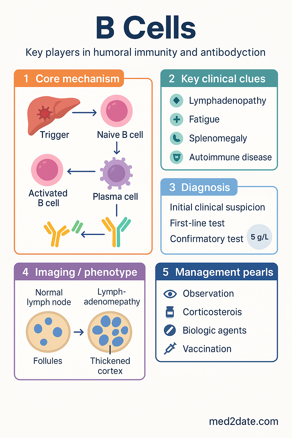
- Activated B cells enter germinal centres where somatic hypermutation (AID-mediated) and class-switch recombination refine antibody affinity and isotype.
- Differentiation yields short-lived plasmablasts (extrafollicular) and long-lived plasma cells (germinal centre-derived) plus memory B cells that provide rapid secondary responses.
- B cell disorders encompass immunodeficiency (XLA, CVID), autoimmunity (SLE, rheumatoid arthritis), and malignancy (CLL, DLBCL, ALL).
- Anti-CD20 therapy (rituximab, ocrelizumab) depletes B cells and is used in lymphoma, rheumatoid arthritis, MS, and ANCA vasculitis; monitor immunoglobulin levels during prolonged use.
- Australian patients on anti-CD20 agents should complete all scheduled vaccinations ≥4 weeks prior to initiation; live vaccines are contraindicated during therapy and for 6–12 months post-treatment.
- Aboriginal and Torres Strait Islander peoples experience higher rates of rheumatic heart disease (antibody-mediated pathology) and may face barriers to specialist immunology referral in remote regions.
- PBS-listed B cell–targeting agents include rituximab (Authority Required for RA, lymphoma, pemphigus), ocrelizumab (Authority Required for MS), and belimumab (Authority Required for SLE).
Introduction & Australian Context
B cells are a subset of lymphocytes responsible for antibody-mediated (humoral) immunity. They originate from haematopoietic stem cells in the bone marrow, where they undergo a tightly regulated programme of gene rearrangement, selection, and maturation before emigrating to secondary lymphoid organs as naïve mature B cells. Upon encountering cognate antigen, B cells activate, proliferate, and differentiate into antibody-secreting plasma cells and memory B cells.
In Australia, disorders of B cell number or function span primary immunodeficiency (affecting approximately 1 in 10,000–50,000 Australians), autoimmune disease (rheumatoid arthritis prevalence ~2%, SLE ~0.05–0.1%), and B cell malignancies (non-Hodgkin lymphoma is the 6th most common cancer nationally, with ~6,300 new diagnoses annually per AIHW data). B cell–depleting therapies are among the most commonly prescribed immunomodulators through the Pharmaceutical Benefits Scheme (PBS).
Understanding B cell biology is foundational to rational use of targeted therapies including anti-CD20 monoclonal antibodies, BTK inhibitors, and BAFF/BLyS antagonists now listed on the PBS for specific indications.
B Cell Development
Haematopoietic Origin & Bone Marrow Maturation
B lymphopoiesis begins in the foetal liver during embryonic development and shifts to the bone marrow after birth, where it continues throughout life albeit declining with age. The process is orchestrated by transcription factors (E2A, EBF1, PAX5) and cytokine signals, principally IL-7 in humans.
| Stage | Key Molecular Events | Surface Markers |
|---|---|---|
| Pro-B cell | D-J rearrangement of Ig heavy chain (IGH); RAG-1/RAG-2 active | CD34+, CD10+, CD19+, CD79a+ |
| Pre-B cell | V-DJ rearrangement complete; μ heavy chain expressed with surrogate light chain (VpreB + λ5) as pre-BCR | CD19+, CD10+, cytoplasmic μ+, pre-BCR+ |
| Immature B cell | V-J rearrangement of light chain (κ then λ); surface IgM expressed | CD19+, IgM+, IgD− |
| Transitional B cell | Emigration from marrow to spleen; BAFF-dependent survival | CD19+, IgM++, CD24++, CD38++ |
| Mature naïve B cell | Co-expression of IgM and IgD via alternative mRNA splicing; quiescent, antigen-inexperienced | CD19+, CD20+, IgM+, IgD+, CD27− |
V(D)J Recombination
The extraordinary diversity of the B cell receptor repertoire (~1011 unique specificities) is generated by somatic recombination of variable (V), diversity (D), and joining (J) gene segments. The recombination-activating genes RAG-1 and RAG-2 recognise recombination signal sequences (RSS) flanking each segment and introduce double-strand breaks, which are resolved by non-homologous end-joining (NHEJ) machinery including Artemis, DNA-PKcs, Ku70/Ku80, and XRCC4/ligase IV.
Central Tolerance
Immature B cells in the marrow undergo negative selection to remove autoreactive clones:
- Receptor editing: Secondary light-chain gene rearrangement to alter specificity (preferred mechanism).
- Clonal deletion: Apoptosis if receptor editing fails to abolish self-reactivity.
- Anergy: Functional silencing of moderately self-reactive cells that emigrate but are non-responsive.
Failure of central tolerance contributes to autoimmune disease; in SLE, evidence of defective receptor editing has been demonstrated in Australian patient cohorts.
B Cell Receptor Structure
Membrane Immunoglobulin
The antigen-binding component of the BCR is membrane immunoglobulin (mIg), comprising two identical heavy chains and two identical light chains linked by disulphide bonds forming a Y-shaped molecule. Each chain contains a variable (V) domain responsible for antigen recognition and constant (C) domains that define isotype and effector function.
| Component | Structure | Function |
|---|---|---|
| Heavy chain (membrane form) | VH + 3–4 CH domains + transmembrane anchor + cytoplasmic tail | Determines isotype (μ, δ, γ, α, ε); transmembrane anchor retains Ig in membrane |
| Light chain (κ or λ) | VL + CL domain | Contributes to antigen-binding site; κ:λ ratio ~2:1 in humans |
| Igα (CD79a) / Igβ (CD79b) | Disulphide-linked heterodimer; each chain has one extracellular Ig-like domain, transmembrane region, and ITAM-containing cytoplasmic tail | Signal transduction: ITAM phosphorylation by Src-family kinases (Lyn, Fyn, Blk) initiates downstream signalling |
Antigen-Binding Site
The antigen-binding site (paratope) is formed by six complementarity-determining regions (CDRs) — three from the heavy chain (CDR-H1, CDR-H2, CDR-H3) and three from the light chain (CDR-L1, CDR-L2, CDR-L3). CDR-H3, encoded by the V-D-J junction, is the most hypervariable and often the dominant contributor to antigen specificity.
Co-Receptors & Accessory Molecules
BCR signalling is modulated by co-receptors that lower the activation threshold or provide inhibitory signals:
- CD19/CD21/CD81 complex: CD21 (complement receptor 2, CR2) binds C3d-opsonised antigen, dramatically lowering the BCR activation threshold (up to 1,000–10,000 fold).
- CD22 (Siglec-2): Inhibitory receptor containing ITIMs; recruits SHP-1 phosphatase to dampen signalling.
- FcγRIIB (CD32B): Inhibitory Fc receptor; co-crosslinking with BCR by immune complexes triggers ITIM-mediated apoptosis or anergy — a key peripheral tolerance mechanism.
Activation Mechanisms
T-Dependent Activation
Protein antigens require T cell help for optimal B cell responses. The process involves:
T-Independent Activation
| Type | Antigen Examples | Mechanism | Clinical Relevance |
|---|---|---|---|
| TI-1 | LPS, CpG DNA | Polyclonal B cell activation via TLR4/TLR9 at high dose; antigen-specific at low dose | Adjuvant effect in vaccines; B-1 cell responses |
| TI-2 | Capsular polysaccharides (e.g., pneumococcal, meningococcal) | Extensive BCR crosslinking by repetitive epitopes; marginal zone B cells respond | Children <2 years have poor TI-2 responses (immature marginal zone); conjugate vaccines circumvent this by recruiting T cell help |
Somatic Hypermutation & Affinity Maturation
Within germinal centres, activation-induced cytidine deaminase (AID) introduces point mutations in the variable-region genes of rearranged Ig at a rate ~106 times the background somatic mutation rate. B cells bearing BCRs with higher affinity for antigen are preferentially rescued from apoptosis by Tfh cells (positive selection); low-affinity and autoreactive clones undergo apoptosis (negative selection). This iterative process, termed affinity maturation, progressively increases serum antibody affinity over the course of an immune response.
Class-Switch Recombination
AID also mediates class-switch recombination (CSR), a deletional recombination event between switch (S) regions upstream of each constant-region gene. CSR changes the heavy-chain constant region (and therefore antibody isotype) without altering antigen specificity. Deficiency of AID causes hyper-IgM syndrome type 2 (HIGM2), characterised by elevated IgM, absent IgG/IgA/IgE, recurrent sinopulmonary infections, and lymphoid hyperplasia.
Differentiation to Plasma Cells
Plasmablast & Plasma Cell Differentiation Pathways
Activated B cells differentiate into antibody-secreting cells via two principal routes:
Activated B cells differentiate rapidly (within 2–3 days) into short-lived plasmablasts that secrete low-affinity, mainly IgM antibodies. Provides early protection before the germinal centre response matures (~7–10 days). Driven by signals including BAFF, APRIL, and IL-21. Short-lived plasma cells undergo apoptosis within weeks.
B cells that successfully undergo SHM and positive selection in the GC light zone differentiate into: (1) long-lived plasma cells that home to the bone marrow survival niche (CXCL12/CXCR4, BAFF/APRIL/BCMA, IL-6) and secrete high-affinity class-switched antibodies for months to decades; and (2) memory B cells that persist in a quiescent state and mount rapid, high-affinity secondary responses upon re-encountering antigen.
Transcriptional Regulation
Plasma cell identity is controlled by a transcriptional network:
- PAX5, BCL-6, Bach2: Maintain B cell identity and GC phenotype; suppress plasma cell differentiation.
- BLIMP-1 (PRDM1): Master regulator of plasma cell differentiation; represses PAX5, CIITA (MHC II), and AID.
- IRF4 (high levels): Induces BLIMP-1; essential for plasma cell commitment.
- XBP-1: Drives the unfolded protein response (UPR) to expand the endoplasmic reticulum, enabling massive immunoglobulin secretion (up to ~10,000 molecules/second per plasma cell).
Memory B Cells
Memory B cells express high-affinity, class-switched BCRs and reside in secondary lymphoid tissues, bone marrow, and peripheral blood. Upon antigen re-encounter, they rapidly differentiate into plasmablasts without requiring the full germinal centre programme. Surface marker phenotype: CD19+, CD20+, CD27+, IgG+ or IgA+ (class-switched). The presence of somatic hypermutations in their Ig genes is a hallmark distinguishing them from naïve B cells.
B Cell Subsets & Functional Diversity
| Subset | Location | Key Features | Primary Function |
|---|---|---|---|
| Follicular (FO) B cells | B cell follicles of spleen and lymph nodes | CD21++, CD23++; T-dependent responses | Germinal centre formation; high-affinity class-switched antibodies |
| Marginal zone (MZ) B cells | Splenic marginal zone | CD21+, CD23−; T-independent responses; rapid IgM secretion | First-line defence against blood-borne encapsulated bacteria |
| B-1 cells | Peritoneal and pleural cavities | CD5+ (B-1a) or CD5− (B-1b); self-renewing; germline-encoded BCRs | Natural antibodies (IgM); innate-like responses to TI antigens |
| Regulatory B cells (Breg) | Various tissues | IL-10–secreting; CD19+, CD24++, CD38++ or CD19+, CD25+, CD71+ | Suppress excessive inflammation; maintain tolerance |
Clinical Relevance — B Cell Disorders
B Cell Immunodeficiencies
| Disorder | Genetic Defect | B Cell Findings | Presentation |
|---|---|---|---|
| X-linked agammaglobulinaemia (XLA, Bruton's) | BTK (Bruton tyrosine kinase) | Absent B cells (<1%); pre-B cell arrest | Males; recurrent sinopulmonary infections from 6–12 months (post-maternal IgG decline) |
| Common variable immunodeficiency (CVID) | Heterogeneous (ICOS, TACI, BAFF-R, CD19) | Low or normal B cell numbers; impaired differentiation to plasma cells; hypogammaglobulinaemia | Adolescents/adults; recurrent infections, autoimmunity, granulomatous disease, increased lymphoma risk |
| Hyper-IgM syndromes | CD40LG (X-linked), CD40, AID, UNG | Elevated IgM; absent IgG, IgA, IgE; defective CSR | Recurrent infections; opportunistic infections (PJP, Cryptosporidium) in CD40L/CD40 defects |
B Cell Malignancies
B cell malignancies are classified by the developmental stage at which transformation occurs:
- Precursor B-ALL: Most common childhood cancer; CD10+, CD19+, TdT+; translocations include t(12;21) ETV6-RUNX1 (good prognosis) and t(9;22) BCR-ABL1 (poor prognosis without TKI therapy).
- CLL/SLL: Most common adult leukaemia in Australia; mature B cells co-expressing CD5 and CD23; smudge cells on blood film; prognostic factors include IGHV mutation status, del(17p)/TP53 mutation, and ZAP-70 expression.
- Diffuse large B cell lymphoma (DLBCL): Most common aggressive NHL; R-CHOP (rituximab, cyclophosphamide, doxorubicin, vincristine, prednisolone) is first-line; PBS-subsidised rituximab.
- Follicular lymphoma: Indolent GC-derived lymphoma; t(14;18) BCL2-IGH; watch-and-wait appropriate for asymptomatic low-burden disease.
B Cell–Mediated Autoimmunity
B cells contribute to autoimmunity through autoantibody production, antigen presentation to autoreactive T cells, and pro-inflammatory cytokine secretion (TNF-α, IL-6, lymphotoxin). Key examples in Australian clinical practice:
- Systemic lupus erythematosus (SLE): Anti-dsDNA, anti-Smith antibodies; B cell hyperactivity; belimumab (anti-BAFF) PBS-authorised.
- Rheumatoid arthritis (RA): Rheumatoid factor (RF), anti-CCP antibodies; rituximab PBS-authorised for methotrexate-refractory disease.
- ANCA-associated vasculitis: Rituximab now first-line for severe GPA/MPA per RACP/ARA guidelines; PBS Authority Required.
- Multiple sclerosis: Ocrelizumab (anti-CD20) PBS-authorised for relapsing and primary progressive MS.
Investigations
B Cell–Targeting Therapies
Special Populations
Aboriginal and Torres Strait Islander Health
📚 References
- 1. Murphy K, Weaver C. Janeway's Immunobiology. 10th ed. New York: Garland Science; 2022. Chapters 7–8: B cell development, activation and differentiation.
- 2. Pieper K, Grimbacher B, Eibel H. B-cell biology and development. J Allergy Clin Immunol. 2013;131(4):959–971.
- 3. Tangye SG, Ma CS, Brink R, et al. The good, the bad and the ugly — TFH cells in human health and disease. Nat Rev Immunol. 2013;13(6):412–426.
- 4. Australian Institute of Health and Welfare (AIHW). Cancer data in Australia. Canberra: AIHW; 2024. Cat. no. CAN 139.
- 5. Australasian Society of Clinical Immunology and Allergy (ASCIA). Primary Immunodeficiency (PID) Guide for Health Professionals. ASCIA, 2023. Available at: https://www.allergy.org.au
- 6. National Health and Medical Research Council (NHMRC). The Australian Immunisation Handbook. 12th ed. Canberra: Australian Government Department of Health; updated 2024. Available at: https://immunisationhandbook.health.gov.au
- 7. Royal Australasian College of Physicians (RACP). Position statement on rituximab use in ANCA-associated vasculitis. Sydney: RACP; 2022.
- 8. Australian Rheumatology Association (ARA). ARA clinical guidance for the management of rheumatoid arthritis. 2023. Available at: https://www.rheumatology.org.au
- 9. Pharmaceutical Benefits Scheme (PBS). PBS Schedule of Pharmaceutical Benefits. Australian Government Department of Health. Available at: https://www.pbs.gov.au. Accessed 2024.
- 10. RHDAustralia (RHD Australia). The 2020 Australian guideline for prevention, diagnosis and management of acute rheumatic fever and rheumatic heart disease. 3rd ed. Menzies School of Health Research; 2020.
- 11. Warnatz K, Salzer U, Rizzi M, et al. B-cell activating factor receptor deficiency is associated with an adult-onset antibody deficiency syndrome in humans. Proc Natl Acad Sci USA. 2009;106(33):13945–13950.
- 12. Smith KGC, Light A, O'Reilly LA, et al. bcl-2 transgene expression inhibits apoptosis in the germinal centre and reveals differences in the selection of memory B cells and bone marrow antibody-forming cells. J Exp Med. 2000;191(3):475–484.
- 13. Australian Commission on Safety and Quality in Health Care (ACSQHC). National Safety and Quality Health Service Standards. 2nd ed. Sydney: ACSQHC; 2021.