📋 Key Information Summary
- Immunoglobulins (antibodies) are Y-shaped glycoproteins composed of two heavy chains and two light chains linked by disulphide bonds.
- Five immunoglobulin classes exist: IgG (most abundant, crosses placenta), IgA (mucosal defence), IgM (first responder), IgD (B-cell receptor), IgE (allergy and parasitic defence).
- IgG has four subclasses (IgG1–4); IgA has two (IgA1, IgA2); subclass deficiencies may cause clinical immunodeficiency despite normal total Ig levels.
- Effector functions include neutralisation, opsonisation, complement activation (classical pathway), antibody-dependent cellular cytotoxicity (ADCC), and mast-cell degranulation.
- Intravenous immunoglobulin (IVIg) is indicated for primary immunodeficiency, immune thrombocytopenia (ITP), Kawasaki disease, Guillain-Barré syndrome, chronic inflammatory demyelinating polyneuropathy (CIDP), and specific autoimmune conditions.
- Subcutaneous immunoglobulin (SCIg) offers equivalent efficacy to IVIg for antibody-replacement therapy with superior steady-state IgG levels and fewer systemic adverse effects.
- IVIg dose varies by indication: 0.4 g/kg/month for replacement; 1–2 g/kg (often over 2–5 days) for immunomodulation.
- Common adverse effects include headache, flushing, myalgia, and rigors; serious risks include anaphylaxis in IgA-deficient patients with anti-IgA antibodies, thromboembolic events, and aseptic meningitis.
- Australian blood-plasma supply is managed by Lifeblood (Australian Red Cross) and CSL Behring; IVIg products available in Australia include Intragam P, Privigen, Flebogamma, and Kiovig.
- In Australia, IVIg and SCIg are funded under the National Blood Agreement; prescribing requires compliance with the National Immunoglobulin Governance Programme (NIGP) and State/Territory Blood Management Committees.
- Aboriginal and Torres Strait Islander peoples have higher rates of infections that may benefit from immunoglobulin therapy but face barriers to specialist referral, plasma-donation infrastructure, and regular infusion access.
- Monoclonal antibodies (therapeutic mAbs) are immunoglobulin-derived biologics and are distinct from pooled immunoglobulin products; they target specific antigens (e.g., rituximab targets CD20).
Introduction & Australian Context
Immunoglobulins (antibodies) are glycoproteins produced by terminally differentiated B lymphocytes (plasma cells) that specifically bind antigens, thereby mediating the effector functions of humoral immunity. Each immunoglobulin molecule comprises two identical heavy chains and two identical light chains arranged in a Y-shaped quaternary structure linked by inter-chain disulphide bonds.
In Australia, immunoglobulin products are classified as biological medicines under the oversight of the Therapeutic Goods Administration (TGA). The supply of plasma-derived and recombinant immunoglobulin products is governed by the National Blood Agreement between the Commonwealth, State, and Territory governments. Australian Red Cross Lifeblood collects approximately 750 000 plasma donations annually, yet domestic demand for immunoglobulin continues to grow at 5–8 % per year, driven by expanding indications for immunomodulation.
The National Immunoglobulin Governance Programme (NIGP) provides a framework for evidence-based, appropriate use of immunoglobulin products. Prescribers must comply with NIGP criteria and obtain approval through their State or Territory Blood Management Committee before initiating immunoglobulin therapy, except in life-threatening emergencies.
This guideline provides a comprehensive overview of immunoglobulin structure, classification, effector functions, and clinical applications relevant to Australian primary care and specialist practice.
Structure — Heavy & Light Chains
Basic Immunoglobulin Architecture
Every immunoglobulin molecule is a heterotetramer consisting of:
- Two identical heavy (H) chains — approximately 50–70 kDa each. The heavy-chain constant region determines the immunoglobulin class (isotype): γ for IgG, α for IgA, μ for IgM, δ for IgD, ε for IgE.
- Two identical light (L) chains — approximately 25 kDa each. Two light-chain isotypes exist: kappa (κ) and lambda (λ). In humans, the normal κ:λ ratio is approximately 2:1.
Domain Structure
Each chain folds into globular domains of approximately 110 amino acids, stabilised by an intra-chain disulphide bond:
- Variable domains (VH and VL) — located at the amino-terminal ends; together they form the antigen-binding site (paratope). Hypervariable regions (complementarity-determining regions, CDR1–3) within the variable domains confer antigen specificity.
- Constant domains (CH1–3 or CH1–4, and CL) — located carboxy-terminal to the variable domains; determine isotype, mediate effector functions, and interact with Fc receptors and complement.
Hinge Region
In IgG, IgA, and IgD, a flexible hinge region between CH1 and CH2 allows the two Fab arms to adopt variable angles, enabling bivalent binding to antigens of differing spacing. IgM and IgE lack a classical hinge region; instead, an additional constant domain (CH4) provides structural rigidity.
Fab and Fc Fragments
Papain digestion cleaves IgG above the hinge to yield:
- Fab (fragment antigen-binding) — one VH–CH1 paired with one VL–CL; monovalent antigen binding.
- Fc (fragment crystallisable) — the paired CH2 and CH3 (or CH3–4 in IgM/IgE) domains; mediates complement activation, Fc-receptor binding, neonatal Fc receptor (FcRn) binding, and determines half-life.
Immunoglobulin Classes
| Property | IgG | IgA | IgM | IgD | IgE |
|---|---|---|---|---|---|
| Serum concentration (adult) | 7.0–16.0 g/L (≈75 % of total Ig) | 0.8–4.5 g/L (≈15 %) | 0.5–2.0 g/L (≈8 %) | Trace (<0.03 g/L) | Trace (<0.00005 g/L) |
| Heavy chain | γ (gamma) | α (alpha) | μ (mu) | δ (delta) | ε (epsilon) |
| Structure | Monomer | Dimer (+ J chain + SC) | Pentamer (+ J chain) | Monomer | Monomer |
| Half-life (days) | 21–28 | 5–6 (serum) | 5 | 2.8 | 2 |
| Placental transfer | Yes (via FcRn) | No | No | No | No |
| Complement activation (classical) | Yes (IgG1, IgG3 > IgG2) | No | Yes (very potent) | No | No |
| Primary role | Secondary/memory response; opsonisation; ADCC; neonatal passive immunity | Mucosal defence (secretory IgA) | Primary response; natural antibodies; BCR | B-cell receptor; B-cell maturation | Type I hypersensitivity; anti-helminthic defence |
IgG Subclasses
IgG is divided into four subclasses with distinct effector profiles:
| Subclass | % of IgG | Half-life (days) | Complement | FcγR binding | Clinical significance |
|---|---|---|---|---|---|
| IgG1 | 60–70 % | 21 | Strong | Strong (FcγRI, FcγRII, FcγRIII) | Most abundant; key opsonin; deficiency causes recurrent sinopulmonary infections |
| IgG2 | 20–30 % | 21 | Moderate | Weak | Anti-polysaccharide responses (pneumococcus, Haemophilus); deficiency → encapsulated organism infections |
| IgG3 | 5–8 % | 7 | Very strong | Very strong | Short half-life due to hinge-region polymorphism; potent pro-inflammatory subclass |
| IgG4 | 3–4 % | 21 | None | Weak | Anti-inflammatory; undergoes Fab-arm exchange; elevated in IgG4-related disease |
IgA Subclasses
IgA1 predominates in serum, whereas IgA2 is more abundant at mucosal surfaces (resistant to bacterial proteases). Secretory IgA (sIgA) is a dimer of IgA monomers linked by a J chain and bound to the secretory component (SC) derived from the polymeric immunoglobulin receptor (pIgR). sIgA is the dominant immunoglobulin in breast milk, saliva, tears, and gut luminal secretions.
Effector Functions
1. Neutralisation
Antibodies bind to pathogen surface molecules (e.g., viral spike proteins, bacterial toxins) and physically block attachment to host-cell receptors. This is the primary mechanism of protection for most viral vaccines, including the influenza and SARS-CoV-2 vaccines used in Australia's National Immunisation Programme (NIP).
2. Opsonisation and Phagocytosis
IgG coating of pathogens (opsonisation) facilitates recognition by Fcγ receptors (FcγRI/CD64, FcγRII/CD32, FcγRIII/CD16) on macrophages and neutrophils, enhancing phagocytosis. IgG1 and IgG3 are the most efficient opsonins due to their strong FcγR binding and long hinge regions.
3. Complement Activation (Classical Pathway)
IgM (pentameric) and IgG (IgG1, IgG3) activate the classical complement pathway by binding C1q to their Fc regions. This leads to:
- C3b deposition → enhanced opsonisation
- C3a and C5a release → anaphylatoxin-mediated inflammation and chemotaxis
- C5b–9 membrane attack complex (MAC) formation → direct lysis of susceptible organisms (especially Neisseria species)
4. Antibody-Dependent Cellular Cytotoxicity (ADCC)
IgG bound to target-cell surfaces engages FcγRIII (CD16) on natural killer (NK) cells, triggering release of perforin and granzymes. ADCC is a critical mechanism of tumour-cell killing in cancer immunotherapy (e.g., rituximab-mediated depletion of CD20⁺ B cells in lymphoma).
5. Mast-Cell and Basophil Degranulation
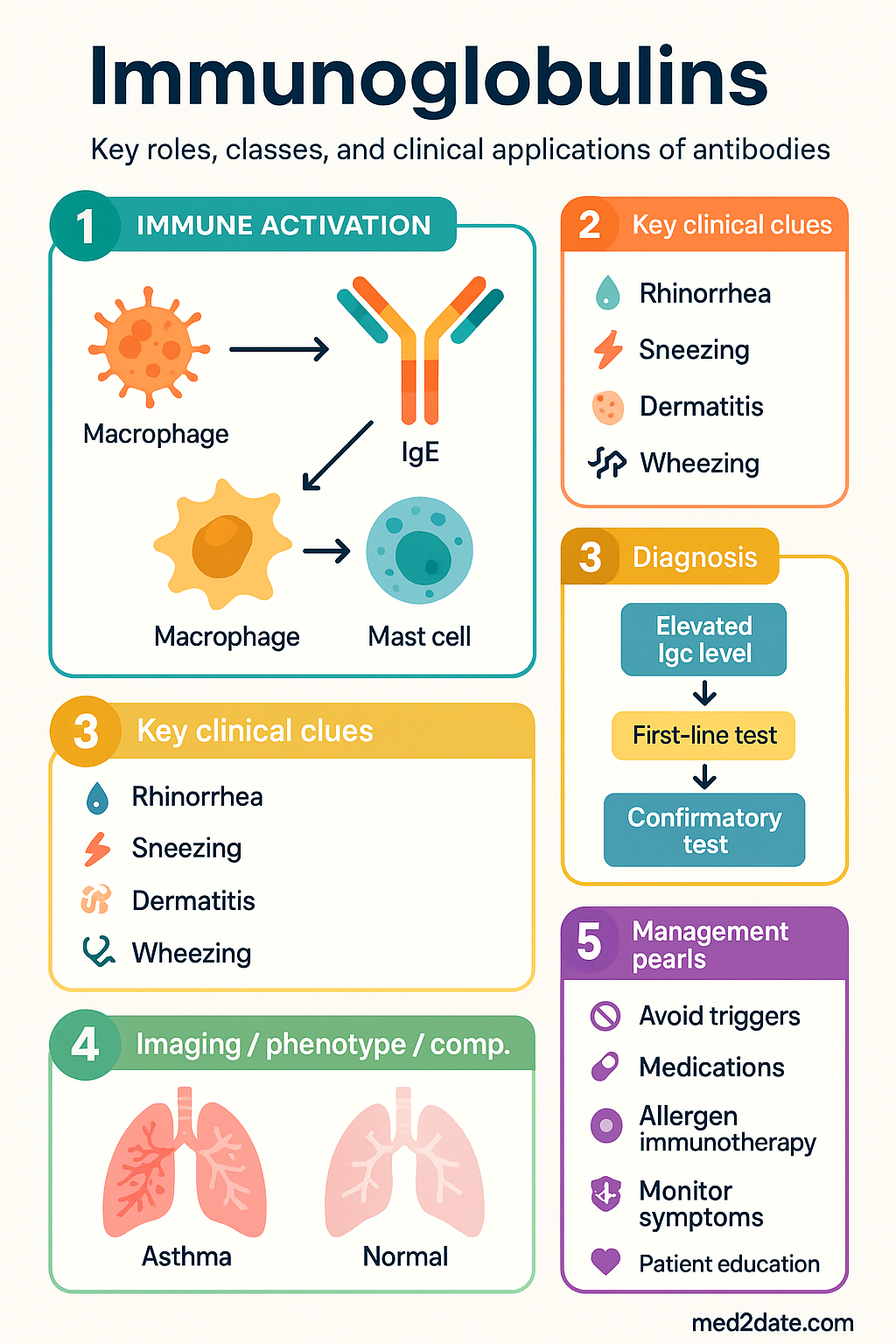
IgE binds with very high affinity (Kd ≈ 10⁻¹⁰ M) to FcεRI on mast cells and basophils. Cross-linking of surface IgE by multivalent allergens triggers degranulation, releasing histamine, leukotrienes, and prostaglandins — the basis of type I hypersensitivity (allergic rhinitis, asthma, anaphylaxis).
6. Neonatal Fc Receptor (FcRn) Recycling
FcRn in vascular endothelium and syncytiotrophoblast binds the Fc region of IgG at acidic pH (endosome), rescuing it from lysosomal degradation and recycling it to the cell surface. This mechanism:
- Extends the IgG half-life to 21–28 days (longest of all immunoglobulin classes).
- Mediates active transplacental transfer of maternal IgG to the foetus, particularly in the third trimester, providing passive neonatal immunity for the first 3–6 months of life.
Clinical Applications
A. Immunoglobulin Replacement Therapy
Indicated for patients with primary or secondary immunodeficiency characterised by impaired antibody production.
B. Immunomodulatory Applications
High-dose immunoglobulin exerts immunomodulatory effects via Fc-receptor saturation, anti-idiotypic antibody-mediated neutralisation of autoantibodies, cytokine modulation, and complement scavenging.
C. IVIg Products Available in Australia
| Product | Manufacturer | Concentration | IgA content | Route |
|---|---|---|---|---|
| Intragam P | CSL Behring | 6 % (60 mg/mL) | ≤ 20 µg/mL | IV |
| Privigen | CSL Behring | 10 % (100 mg/mL) | ≤ 25 µg/mL | IV |
| Flebogamma 10 % DIF | Grifols | 10 % (100 mg/mL) | ≤ 50 µg/mL | IV |
| Kiovig | Takeda / Baxalta | 10 % (100 mg/mL) | ≤ 10 µg/mL | IV |
| Hizentra | CSL Behring | 20 % (200 mg/mL) | ≤ 50 µg/mL | SC |
| Cuvitru | Takeda / Baxalta | 20 % (200 mg/mL) | ≤ 10 µg/mL | SC |
| Subcuvia | CSL Behring | 16 % (160 mg/mL) | ≤ 320 µg/mL | SC |
D. Adverse Effects & Safety Monitoring
| Adverse Effect | Frequency | Management |
|---|---|---|
| Headache, myalgia, chills, flushing | Common (5–15 %) | Slow rate; paracetamol; IV fluids |
| Aseptic meningitis | Uncommon (< 5 %) | Stop infusion; LP if diagnostic uncertainty; supportive care; consider alternative product |
| Anaphylaxis (IgA-deficient patients) | Rare (< 1 %) | IM adrenaline 0.01 mg/kg (max 0.5 mg adults); switch to IgA-depleted product |
| Thromboembolism (DVT, PE, stroke, MI) | Rare (< 1 %) | Risk mitigation: adequate hydration, slow infusion rate, avoid concurrent high-dose IV corticosteroids |
| Haemolytic anaemia (anti-A/anti-B) | Uncommon | Monitor FBC and haptoglobin post-infusion; ABO-compatible products where available |
| Renal impairment (sucrose-containing products) | Rare | Avoid sucrose-stabilised formulations in CKD; monitor eGFR; use osmotic-nephrosis-safe products (e.g., Intragam P, Privigen) |
| Transfusion-transmission infection | Extremely rare | Australian products undergo multiple virucidal steps (pasteurisation, low-pH incubation, solvent/detergent, nanofiltration) |
E. Therapeutic Monoclonal Antibodies (Selected)
Recombinant monoclonal antibodies are engineered immunoglobulin-derived biologics. Selected examples relevant to Australian practice include:
| Monoclonal antibody | Target | Indication | PBS status |
|---|---|---|---|
| Rituximab (MabThera®) | CD20 | B-cell lymphoma, CLL, RA, ANCA vasculitis | ✔ PBS General Benefit |
| Trastuzumab (Herceptin®) | HER2 | HER2⁺ breast cancer | ✔ PBS General Benefit |
| Infliximab (Remicade®) | TNF-α | Crohn disease, ulcerative colitis, RA, psoriasis | ✔ PBS Restricted Benefit |
| Omalizumab (Xolair®) | IgE (Cε3 domain) | Severe allergic asthma, chronic spontaneous urticaria | ✔ PBS Restricted Benefit |
| Dupilumab (Dupixent®) | IL-4Rα | Moderate–severe atopic dermatitis, asthma, CRSwNP | ✔ PBS Restricted Benefit |
| Nivolumab (Opdivo®) | PD-1 | Melanoma, NSCLC, RCC, others | ✔ PBS Restricted Benefit |
F. Diagnostic Immunoglobulin Assessment
Immunoglobulin quantification is performed by nephelometry or turbidimetry (RCPA-accredited laboratories). Key indications for testing include recurrent infections, suspected immunodeficiency, paraproteinaemia, and autoimmune disease monitoring.
G. Vaccination Considerations
Patients receiving immunoglobulin replacement therapy have impaired vaccine antibody responses during and for several months after infusion. Key Australian recommendations include:
- Live vaccines (MMR, varicella, yellow fever) — contraindicated during immunoglobulin therapy and for ≥ 8 months after the last dose (longer interval for high-dose immunomodulatory IVIg).
- Inactivated vaccines — can be administered but antibody responses may be blunted; consider checking post-vaccination titres (e.g., anti-pneumococcal IgG) 4–6 weeks after vaccination.
- Influenza vaccine — recommended annually per NIP schedule regardless of immunoglobulin therapy.
- COVID-19 vaccine — recommended per ATAGI guidance; immunoglobulin therapy does not alter the vaccine schedule.
Special Populations
Aboriginal and Torres Strait Islander Health Considerations
📚 References
- 1. Australian Government Department of Health and Aged Care. National Immunoglobulin Governance Programme: Clinical Criteria for Immunoglobulin Use in Australia. 3rd edn. Canberra: Commonwealth of Australia; 2015 (updated 2022).
- 2. Australian Technical Advisory Group on Immunisation (ATAGI). Australian Immunisation Handbook. Australian Government Department of Health and Aged Care, Canberra; 2022. Available at: immunisationhandbook.health.gov.au.
- 3. National Blood Authority Australia. National Blood Agreement. Canberra: NBA; 2003 (revised 2021).
- 4. Royal College of Pathologists of Australasia (RCPA). Immunoglobulin quantification and electrophoresis: RCPA Manual. Sydney: RCPA; 2023.
- 5. Australasian Society of Clinical Immunology and Allergy (ASCIA). Primary Immunodeficiency (PID) Position Statement. Sydney: ASCIA; 2023.
- 6. Ballow M, Cunningham-Rundles C. Intravenous immunoglobulin: clinical applications and safety. J Allergy Clin Immunol. 2023;151(3):612–624.
- 7. Orange JS, Grossman WJ, Navickis RJ, Wilkes MM. Impact of trough IgG on pneumonia incidence in primary immunodeficiency: a meta-analysis of clinical studies. Clin Immunol. 2010;137(1):21–30.
- 8. Perez EE, Orange JS, Bonilla F, et al. Update on the use of immunoglobulin in human disease: a review of evidence. J Allergy Clin Immunol. 2017;139(3S):S1–S46.
- 9. Australian Institute of Health and Welfare (AIHW). Aboriginal and Torres Strait Islander Health Performance Framework 2023 Summary Report. Canberra: AIHW; 2023.
- 10. Schroeder HW Jr, Cavacini L. Structure and function of immunoglobulins. J Allergy Clin Immunol. 2010;125(2 Suppl 2):S41–S52.
- 11. Katz U, Shoenfeld Y, Zandman-Goddard G. Update on intravenous immunoglobulins (IVIg) mechanisms of action and off-label use in autoimmune diseases. Curr Pharm Des. 2011;17(29):3166–3175.
- 12. Hodkinson JP. Considerations for dosing immunoglobulin in obese patients. Immunotherapy. 2019;11(11):967–975.
- 13. Gelfand EW. Intravenous immune globulin in autoimmune and inflammatory diseases. N Engl J Med. 2012;367(21):2015–2025.
- 14. Emergency Care Institute, NSW. Kawasaki Disease: Clinical Practice Guideline. Sydney: ACI; 2022.
- 15. National Blood Authority Australia. Criteria for the Clinical Use of Intravenous Immunoglobulin in Australia. Canberra: NBA; 2012.